Sunday, December 29, 2013

Salivary Mucoceles in Dogs: Cervical, Oral (rannula), and 1 type that causes airway obstruction

Anatomy


Fig. 1
(from: Manfra Marretta S. Dentistry and diseases of the oropharynx, in Birchard & Sherding (editors): Saunders Manual of Small Animal Practice, (3rd ed.). St. Louis. Elsevier, 2006, pp 632-635.)
The mandibular salivary gland is located at the bifurcation of the maxillary and linguofacial veins. (Fig. 1) The duct empties at the sublingual papilla, which is lateral to the most rostral aspect of the frenulum of the tongue. The gland has a well-defined capsule. The sublingual gland has two distinct portions, the monostomatic portion, which is immediately adjacent and rostral to the mandibular gland, and the polystomatic portion, which are small islands of gland dispersed along the salivary duct. The sublingual salivary duct is closely associated with the mandibular duct. The parotid gland is located at the base of the ear canal. Its duct empties adjacent to the upper 4th premolar. This is a wide, but thin gland; not globoid like the mandibular gland. The zygomatic gland is just ventral and medial to the zygomatic arch and the duct empties 1 cm caudal to the parotid duct.

Etiology of mucocele

 A salivary mucocele is the accumulation of saliva outside of the gland or duct system. The mucocele is not a true cyst since it does not have a secretory lining. It can be due to a variety of causes. Trauma to the head or neck that causes injury to either the gland or the duct can cause leakage of saliva. Oral mucocele (ranula) is a reported complication of mandibulectomy in dogs and cats. Infection or inflammation (sialadenitis) may cause enough tissue disruption to allow leakage. Histopathology of salivary tissue removed to treat mucoceles frequently reveals inflammatory changes. Although rare in dogs and cats but more common in humans, calculi can cause obstruction of the salivary duct with subsequent rupture of the duct and leakage. Rarely, neoplasia of the gland can cause disruption of glandular or ductal tissues.
Mucoceles are classified by their location. The cervical mucocele is the most common. A fluctuant mass is seen in the submandibular region and the leaking gland or duct is either the mandibular or the sublingual. If large, the mass may be mid-cervical or encompass the entire ventral aspect of the neck. An oral mucocele, also called a ranula, is due to leakage from the sublingual gland or duct and the swelling is intra-oral and lateral to the tongue. A pharyngeal mucocele is very similar to cervical but the fluctuant swelling is present in the pharynx, near the tonsil. Like the cervical mucocele, the pharyngeal is due to leakage from either the mandibular or sublingual salivary glands. Finally, the zygomatic mucocele can cause exophthalmus and/or peri-orbital swelling and is due to leakage from the zygomatic salivary gland.

Clinical Signs

Clinical signs of mucoceles vary according to their location.  The cervical mucocele causes a fluctuant, non-painful swelling in the cranial ventral cervical area. (Fig. 2)
Fig. 2: Cervical mucocele in a dog
 
The mass usually lateralizes to the affected side. However, in some cases the mass is directly on midline making it difficult to determine whether the right or left salivary glands are causing the problem. 
The oral mucocele, or rannula, can cause dysphagia, anorexia, excessive salivation, abnormal movements or protrusion of the tongue. (See previous post on Ollie for a case example.) The ranula may be large enough to be visible by the owner. It appears cystic and can be large enough to deviate the tongue to the opposite side.
            Dogs with a pharyngeal mucocele (Fig. 3) can present for inspiratory stertor and dyspnea since the fluid filled mass causes a physical obstruction in the pharynx and upper airway.
Fig. 3: Pharyngeal mucocele (arrow) in a poodle
Difficulty swallowing can also be a sign of pharyngeal mucocele.
A mucocele of the zygomatic salivary gland can cause exophthalmus, divergent strabismus, and a fluctuant swelling in the orbital area.

Diagnosis

            In most cases, salivary mucoceles are not a diagnostic challenge. The animals have a history of an acute or chronic fluctuant swelling that is not painful. Aspiration of the mass typically reveals a straw colored, mucinous fluid that appears ropey when pushed through a needle and syringe onto a slide. (Fig. 4)
Fig. 4: FNA of a mucocele (www.acvs.org)
Cytology of the fluid reveals few cells unless the mucocele is very chronic or infected in which case more evidence of inflammation is seen.
            Diagnostic imaging is usually not necessary but may be indicated in complicated cases or ones in which the presentation is atypical. Ultrasound or computed tomography may be helpful in animals with zygomatic mucoceles to differentiate the mass from neoplasia or foreign body.
            In some animals, the cervical mucocele is difficult to lateralize because it is either directly on midline or very large. Simple methods to determine the affected side are to lay the dog on its back and see to which side the mucocele gravitates. (Fig. 5)
Fig. 5: Placing the dog in dorsal recumbency can help lateralize the mucocele.
Sometimes by pushing medially on the mucocele while doing an oral exam the clinician can see one side of the pharyngeal wall bulge inward. In the rare case where the affected side is still unclear after these manipulations, positive contrast sialography can be performed. Contrast material is injected into the salivary duct and the resultant images studied for evidence of leakage or obstruction.

Treatment

            Regardless of location, the definitive treatment of a salivary mucocele involves removal of the offending salivary glands. Performing only drainage of the mucocele will not result in long-term resolution.
Surgical excision of mandibular and sublingual glands is the treatment of choice for cervical mucoceles. (Fig. 6)
Fig. 6: Excised mandibular (large gland to the left) and sublingual (remaining glands extending
from left to right) salvary glands
The mandibular gland, and the monostomatic and polystomatic portions of the sublingual gland are removed as a unit since their ducts are closely associated. The surgical approach is directly over the mandibular gland that lies just cranial to the bifurcation of the jugular vein into the maxillary and linguofacial veins. The capsule of the mandibular gland is incised to allow dissection and removal of the gland. Dissection then proceeds cranially along the mandibular duct. (Fig. 7)
Fig. 7: Surgical excision of mandibular and sublingual salivary glands in a dog.
Retraction of the digastricus muscle (arrow) facilitates dissection.
The multiple portions of sublingual gland are removed with the mandibular gland and the ducts. Dissect the ducts as far cranially as possible, then ligate and remove the tissues. Take care to avoid injury to the lingual nerve which lies over the salivary duct and serves as the most rostral limit of the gland and duct excision. Place a pen-rose or closed suction drain in the mucocele and close the incision routinely.
                        The etiology of pharyngeal mucoceles is similar to cervical mucoceles since they are also caused by leakage from the mandibular or sublingual glands. Therefore, remove these glands as described under cervical mucoceles. In addition, excise the pharyngeal mucocele via an intra-oral approach. Make an elliptical incision at the base of the mucocele, and excise the redundant mucosa and underlying tissues to be sure that the interior of the mucocele has been exposed. Although marsupialization of the resultant pharyngeal defect has been described, the author simply leaves this incision open to heal by second intention.
                        An oral mucocele, or ranula, is treated by “deroofing” the mucocele, followed by marsupialization. (See previous post on Ollie.) Deroofing is performed by simply removing the mucosa over the dorsal portion of the swelling being sure to expose the inside of the mucocele. Marsupialization is performed by suturing the inner lining of the mucocele to the oral mucosa. This allows the mucocele to remain open and drain into the mouth. Suturing is usually done with an absorbable suture such as Monocryl or PDS. Removal of the mandibular and sublingual salivary glands on the affected side is also be performed to prevent recurrence.
                        The zygomatic mucocele is treated by removal of the zygomatic salivary gland, which is located just ventral to the eye and medial to the zygomatic arch. Removal of the gland can be performed by partial removal of the zygomatic arch, or by ventral orbitotomy. The zygomatic arch can also be temporarily removed to provide exposure, and then reattached with orthopedic wire. Be careful not to injure orbital structures during the dissection.          

Postoperative Care

            Submit removed tissues for histopathology and culture. If a pen-rose drain was left in the mucocele, remove it once drainage becomes minimal. If infection was suspected or documented by positive cultures, treat the dog with appropriate antibiotics for at least 7-10 days. After marsupialization of a ranula, feed the dog soft food for 2-3 weeks, and flush the mouth with water or oral antiseptic lavage after eating.
            Complications after treatment of a salivary mucocele are rare. A seroma can occur due to the dead space created by the mucocele. Conservative treatment with warm compresses is usually effective. Recurrence of mucocele is rare but may indicate incomplete removal of the affected salivary tissue, or that the incorrect gland was removed.

References

Bellenger CR, Simpson DJ. Canine sialocoeles – 60 clinical cases. J of Sm. An. Practice 33:376-380, 1992.
Schmidt GM, Betts CW. Zygomatic salivary mucoceles in the dog. J Am Vet Med Assoc 172:940-942, 1978.
Knecht CD. Diseases of the salivary glands in the dog. Comp Cont Ed II:932-938, 1980
Bartoe JT, Brightman AH, Davidson HJ. Modified lateral orbitotomy for vision-sparing excision of a zygomatic mucocele in a dog. Vet Ophthalmology10:127-131, 2007.
Manfra Marretta S. Dentistry and diseases of the oropharynx, in Birchard & Sherding (editors): Saunders Manual of Small Animal Practice, (3rd ed.). St. Louis. Elsevier, 2006, pp 632-635.
Benjamino K, Birchard SJ, Niles JD, Penrod KD. Pharyngeal mucoceles in dogs: 14 cases. J Am Anim Hosp Assoc. 48(1):31-5, 2012

Tuesday, December 17, 2013

Outcome of Diagnosis and Treatment of "Ollie"

Fig. 1: FNA of the oral mass in Ollie

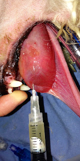
The gross appearance of the mass, its fluctuant nature, and the mucoid fluid that was aspirated (Fig. 1) were all suggestive of a rannula (oral mucocele). This is an accumulation of saliva from the sublingual salivary glands into the submucosal space in the mouth. 

The treatment for Ollie was marsupialization of the rannula (Fig. 2-3), and removal of the mandibular and sublingual salivary glands. (Fig. 4) 
Fig. 2: Completed marsupializaton of the rannula
Fig. 3: Close up of the marsupialized rannula showing the interior of the mucocele.

Marsupialization was performed by first incising over the dorsal aspect of the rannula with a scalpel. The saliva was evacuated from the cavity and the dorsal aspect of the rannula was debrided to make a large opening. The wall of the rannula was sutured to the inner tissue layer with 4-0 Monocryl in a simple continuous pattern. 

The right mandibular and sublingual salivary glands were removed in the standard fashion through a lateral cervical incision directly over the mandibular gland. (Fig. 4)
Fig. 4: Excised mandibular (yellow arrow) and sublingual (red arrows) salivary glands
in Ollie. Note the discolored areas of the sublingual glands. The salivary leakage was
likely originating from these glands.
Ollie was discharged the day after surgery with instructions to feed a soft diet and to flush the mouth with warm water after eating.

Ollie returned in 2 weeks for suture removal. The rannula had resolved and Ollie was eating and drinking normally and doing very well. 

In the next blog we will discuss salivary mucoceles in more detail.

Please post any questions you have about Ollie.

Monday, December 16, 2013

What's Your Diagnosis on "Ollie": A 5 year old Bichon with a huge oral mass?

Fig. 1
Ollie (Fig. 1) is a 5 year old male neutered Bichon who presented with a 5 day history of lethargy, discomfort, and excessive drooling. Oral exam revealed a large, soft mass just lateral to the frenulum of the tongue on the right side. (Fig. 2).
Fig. 2
Fine needle aspirate revealed a tan colored, thick, mucoid fluid. (Fig. 3)
Fig. 3

What is your diagnosis? What other diagnostic tests should be performed?
What is your recommended treatment?

Answer later this week!

Sunday, December 8, 2013

Intestinal surgery quiz


1. True or False: Mortality rate associated with linear foreign bodies is the same as non-linear foreign bodies (such as a corn cob or ball).

2. In an intussuception, the section of intestine that is telescoped inside is called the:
            a. intussusceptum
            b. intussucipiens
            c. intussusinside
            d. dead part
            e. who cares?

3. What is the reported mortality rate for dogs having intestinal leakage after enterotomy or resection and anastomosis:
            a. 10%
            b. 30%
            c. 50%
            d. 75%
            e. 100%

  4. What is the “holding” or strongest histologic layer of the intestine:
a. serosa
b. muscularis
c. submucosa
d. mucosa
e. pseudostratified epithelium

  5. Intestinal anastomotic breakdown usually occurs:
            a. 1-2 days post-operatively
            b. 3-5 days post-operatively
            c. 5-7 days post-operatively
            d. 7-10 days post-operatively
            e. if you don’t sing “Kum-bi-ya”

 6. Which of the following is true about intussuception in dogs and cats:
a.     usually occurs at the ileo-ceco-colic junction in dogs
b.     is frequently not reducible
c.     is usually secondary to some primary disease process
d.     usually occurs in the jejunum in cats, and is associated with inflammatory bowel disease or lymphosarcoma in older cats
e.     all of the above are true

7. Name possible causes of ileus after intestinal surgery.
            a. pain (sympathetic stimulation)
            b. peritonitis
            c. hypokalemia
            d. mechanical obstruction
            e. All of the above

8. Name possible clinical signs of intestinal leakage after resection and anastomosis.
            a. fever
            b. abdominal pain
            c. anorexia
            d. vomiting
            e. all of the above

9. True or False: Linear foreign bodies sometimes cause full thickness perforation of the mesenteric aspect of the intestine.

10.  Which suture is best for closure of the intestine in a dog or cat:
            a. 2-0 catgut
            b. 3-0 polypropylene,  CA 2 needle
            c. 4-0 PDS, AR 1 needle
            d. 4-0 PDS, RB 1 needle
            e. 4-0 stainless steel
           

Answers: F, a, d, c, b, e, e, e, T, d

Thursday, December 5, 2013

4 Ways to Avoid Leaving a Sponge in the Abdomen of a Dog, and Look What Else Someone Left In!

1. Count! Do a sponge count before and after the surgical procedure. If there is a discrepancy, search the surgical site for the sponge. If no sponge is found, obtain a postoperative radiograph to determine if a sponge is retained (see item #2).
 
Fig. 1: Ray-tek 4 X 4 sponges on the left have a radiopaque line in them (the blue material)
Larger laparotomy sponges on the right have a blue radiopaque ribbon on them. 
2. Use Ray-tek sponges. They have linear radiopaque material in them so that the sponge can be easily seen on radiographs. (Fig. 1)
 
Fig. 2: A sponge left in a dog during a spay. Bowel and mesentery are adhered to the sponge
making removal difficult.
3. Never pack an entire sponge in a body cavity. As the sponge absorbs blood and other fluids it blends in with the tissues and becomes almost invisible. (Fig. 2) Always leave part of the sponge outside of the cavity, or attach a forcep to it and keep the forcep outside of the cavity.

4. Use laparotomy sponges. (Fig. 1) These large, very absorbent sponges are a better choice than 4 x 4 sponges for packing off organs in the abdomen or thorax. A blue radiopaque ribbon is attached to them that can be left outside of the body cavity. Having said this, I have seen these left in the abdomen as well. Count them before and after the surgery also.
 
Fig. 3: Abdominal radiograph of a 4 year old female St. Bernard that was
spayed as a puppy.
Sponges are not the only things left in patients. (Fig. 3) The radiograph is a St. Bernard I operated many years ago at the Animal Medical Center in New York City. The dog had been spayed several years earlier. The dog presented to us for vomiting and abdominal pain. On abdominal exploratory, a section of bowel mesentery had become entrapped and strangulated in one of the rings of the forceps. The forceps were rusted and broken. Intestinal resection and anastomosis was necessary.


Establish protocols for all surgical procedures to prevent mistakes. Although they seem tedious, they are necessary to avoid complications and resultant patient morbidity.

Sunday, November 24, 2013

Here Is What Happens When You Leave a Surgical Sponge in the Abdomen

Fig. 1
Fig. 2

Case Report

Penny is a 5-year-old female spayed mixed breed dog that presented with a chronic draining tract on her left flank. (Fig. 1-2) The track had been present for 1 year and had been operated 3 times with no resolution. Each time the exploratory surgery was performed directly over the affected area of the flank attempting to follow the tract to its source. No foreign body or other etiology was found on any of the previous surgeries. Repeated antibiotic therapy would temporarily stop the drainage; cessation of the antibiotic would result in recurrence of the drainage. Other than the tract no significant abnormalities were found on physical examination.

Plain film abdominal radiographs were unremarkable. A positive contrast fistulogram was performed by inserting a Foley catheter into the tract and injecting water-soluble radiographic contrast material.(Fig. 3) Although a foreign body was not outlined it appeared that the contrast entered the abdominal cavity.
Fig. 3

Because of the suspicion of an intra-abdominal foreign body, a ventral midline laparotomy was performed. A large, adherent, firm granuloma was present in the left dorsal caudal abdomen adjacent to the urinary bladder and descending colon and adhered to the left ureter. (Fig. 4) 
Fig. 4: Granuloma in caudal abdomen (arrow)
After tedious blunt and sharp dissection that extended deep into the epaxial muscles, the granuloma was excised. Inside the granuloma was a surgical sponge. (Fig. 5)
Fig. 5: Surgical sponge found in the center of the granuloma.
Penny had an unremarkable recovery from anesthesia and surgery. The tract resolved and did not recur.

The only abdominal surgery Penny had was when she was spayed as a puppy. The sponge was present in her for 5 years.

In the next blog we’ll talk about how to prevent this from happening.

Thursday, November 21, 2013

Dehiscence of the Intestinal Incision: Why does it happen and how can we prevent it?

Dehiscence of an enterotomy of the jejunum in a dog
You have just completed an intestinal anastomosis. The bowel looks healthy, there was minimal contamination, and the remainder of the abdomen looks good. The omentum has been tucked into place around the bowel and you close the abdominal incision.

Now begins the period of postoperative anxiety (for you, not the patient). For the next 3 days every time the dog vomits, spikes a fever, doesn’t eat, or seems painful in the abdomen, your “worry-meter” will go off the charts.  You will ask yourself many questions: did you remove all of the diseased bowel, are your sutures holding, was there another perforation or foreign body that you missed, or is the animal just not healing properly? The source of the anxiety of course is that postoperative intestinal leakage is a devastating complication. One study found a dehiscence incidence of 16% of enterotomies and anastomoses in dogs, with a mortality rate of 74%.1 Dogs with foreign bodies and trauma appeared to be at higher risk for dehiscence in this study. Clinical evidence of peritonitis appeared at about 3 days postoperatively.

Intestinal wound breakdown can also occur after full thickness biopsy.  Eight of 66 dogs died or were euthanized due to dehiscence of intestinal biopsy sites in a British clinical study.2 No specific factors predisposing dogs to biopsy dehiscence were identified in the study.

Pathophysiology

Why do intestinal incisions sometimes breakdown and can we do anything to prevent it? In a study of dogs having intestinal surgery, high risk for dehiscence was found if a dog had 2 of the 3 following clinical factors: pre-existing peritonitis, foreign body, or a serum albumin of less than 2.5g/dl.3 This is valuable information and serves to heighten the surgeon’s awareness of postoperative septic peritonitis in selected dogs or cats that fit these criteria. Monitoring of pre- and postoperative serum albumin levels after gastrointestinal surgery, in addition to routine patient parameters (temperature, abdominal pain, vomiting, etc.), appears warranted.

In addition to these well-documented dehiscence factors, clinical experience tells us that dogs with metastatic intestinal neoplasia are also at higher risk for incisional breakdown. Dogs with abdominal carcinomatosis seem particularly prone to poor healing of their incisions.

Prevention

Prevention of peritonitis secondary to intraoperative contamination is by following good surgical technique: packing off the intestine with abdominal sponges, keeping tissues moist, atraumatic technique, use proper suture materials and patterns, and short surgical time. Abdominal lavage is not indicated in dogs that do not have generalized peritonitis. Local lavage of the affected intestinal segment is fine but do not allow fluid to enter the peritoneal cavity. Abdominal lavage fluid has been found to potentiate peritonitis by distributing bacteria and inhibiting phagocytosis by macophages and neutrophils. Abdominal lavage however is indicated in animals with existing peritonitis.

Prophylactic antibiotics, i.e. those given prior to and during surgery but not postoperatively, are also recommended for gastrointestinal surgery in dogs and cats. However, unless the animal already has septic peritonitis, antibiotics are not continued after surgery because continued administration has no benefit and may actually be detrimental. Unnecessary antibiotic administration leads to development of resistant bacterial species and may mask the early signs of peritonitis making it more difficult to diagnose.
Closed suction drain (Jackson-Pratt) used for abdominal drainage in peritonitis
Clinical signs of postoperative septic peritonitis are: abdominal pain, vomiting, anorexia, fever, neutrophila and left shift on complete blood count, and peritoneal fluid that contains bacteria. Treatment is re-operation, resection of the involved intestinal segment (do not try to just re-suture it), abdominal lavage with copious amounts of sterile saline, and placement of a closed suction drain (e.g. Jackson Pratt). Postoperative care includes fluids and colloids, antibiotics, nutritional support, and careful monitoring.

References

1. Allen DA, Smeak DD, Schertel ER. Prevalence of small intestinal dehiscence and associated clinical factors: a retrospective study of 121 dogs. J Am Anim Hosp Assoc 1992;28:70-76.
2. C J Shales, J Warren, D M Anderson, S J Baines, R A S White. Complications following full-thickness small intestinal biopsy in 66 dogs: a retrospective study. Journal of Small Animal Practice 08/2005; 46(7):317-21.
 3. Ralphs SC, Jessen CR, Lipowitz AJ. Risk factors for leakage following intestinal anastomosis in dogs and cats: 115 cases (1991-2000). J Am Vet Med Assoc 2003;223:73-77

Thursday, November 14, 2013

Case Outcome: German Shepherd With Chronic Diarrhea (Did you get it right?)

Fig. 1: note intraluminal soft tissue mass in the colon in the lateral abdominal radiograph
Fig. 2: Note filling defect in the colon in the barium study

Diagnosis 

Cecal inversion (or cecal intussusception). The cecum is "inside out" and what you see on the surgical photo is the cecal mucosa.

Treatment

Typhlectomy (removal of the cecum) 

After exposing the cecum via a colotomy it was excised at its base and the colotomy closed routinely in one layer. (simple continuous, 4-0 PDS) Michael did well and made a full recovery.

Discussion

Cecal inversion, or ceco-colic intussusception, is a rare disorder that is characterized by hematochezia and occasionally, diarrhea. It is more common in young dogs. A palpable abdominal mass is present and the primary differential diagnoses are foreign body, ileo-ceco-colic intussusception, and neoplasia. Typhlitis, such as that associated with whipworm infestation, may predispose to the condition. Definitive diagnosis is by plain film and contrast radiographs, or abdominal ultrasound


Fig. 3: Pneumocologram in a dog with cecal inversion. Note intraluminal soft
tissue mass.(arrows)  (from: Leah A. Cohn, DVM, PhD, DACVIM; Jimmy C. Lattimer, DVM, MS, DACVR; and Laura D. Dvorak, DVM. What’s your diagnosis? JAVMA, Vol 220, No. 2, pg 169, January 15, 2002.)
Pneumocologram (Fig. 3) or colonoscopy can also be used to confirm the diagnosis. Treatment is surgical removal of the cecum. Removal of the cecum, also called typhlectomy, can be done by first performing a colotomy to exteriorize the cecum, then removing the cecum at its base. Closure of the resultant colonic incision is routine. Alternatively, resection of the ileo-ceco-colic junction can be performed. However, removal of only the cecum, preserving the ileo-ceco-colic valve, is preferable. Attempts to un-invert the cecum are fruitless because of the severe inflammation and edema of the tissue. Be sure to treat associated parasitic infestations.


Tuesday, November 12, 2013

What's Your Diagnosis?: 1.5 yr old German Shepherd with chronic diarrhea (An Unusual Diagnosis!)


Michael

History and Physical Examination:

Michael is a 1 1/2 year old male castrated German Shepherd presented with a history of bloody diarrhea for 2 weeks. Frank blood has been noted in the stool. On physical examination, a mid-abdominal, movable tubular mass was palpated. Pain was elicited on palpation.  Fecal examination revealed whipworms.

Imaging

Review the plain film lateral abdominal radiograph and upper gastrointestinal barium series (Fig 1-2). (The ventro-dorsal projections were not contributory.)
Fig. 1
Fig. 2

A ventral midline exploratory laparotomy was performed, and the ileo-ceco-colic region was exposed. (Fig. 3: Ileum to the right, colon to the left) 
Fig. 3
A colotomy was performed on the antimesenteric aspect of the proximal colon and a structure was extruded from the colonic lumen. (Fig. 4)
Fig. 4
What abnormalities are seen on the radiographs? (Fig 1-2)

What is the structure that came out of the colon? (Fig. 4)

What is the diagnosis?

What is the treatment and prognosis?

Answers coming soon!

Sunday, November 3, 2013

Are Some Doctors Born To Be Surgeons, Or Can It Be Learned?


I was a 2nd-year veterinary student, and it was my turn to be the primary surgeon in our surgical lab course. The procedure to be performed was a splenectomy, and it was on a live dog. (Many years ago, shelter or purpose-bred dogs were used for various surgical procedures in teaching labs like this. Today surgical laboratories in many veterinary schools do spay and neuter shelter dogs that are then adopted.)


I was nervous about this lab days in advance. I was a surgery “virgin”; it was my first time as the primary. I was afraid I would make a mistake that hurt the dog and ruined my confidence. I knew these labs were a “rite of passage” for vet students in their journey to becoming a doctor, and I didn’t want to fail. My quest to be a vet began when I was ten years old; I didn’t want it to end prematurely because I couldn’t successfully do surgery.


The surgery began fine but quickly deteriorated. I made the initial incision and opened the abdominal cavity, but I was afraid to pull the spleen out. I thought if I were too rough, it would rupture, and the dog would bleed to death. (Splenic rupture can lead to life-threatening hemorrhage, so there was some logic to my hysteria.) After about an hour, which felt like an entire day, I finally got the spleen exteriorized. It was enormous. The anesthetic drugs had caused severe splenomegaly; to me, it looked like a gigantic purple throbbing alien ready to explode and create an epic disaster.


I had to identify the splenic blood vessels and get them ligated to remove the scary organ before it ruined my life. For those who have never done a splenectomy, about 16,000 little blood vessels must be tied off. I thought I had to dissect and ligate every tiny vessel individually, so I started doing that. The minutes and hours ticked away. After a while, the student doing anesthesia (who probably still hates me to this day) rudely announced that the dog was going into shock; pale mucous membranes and rapid heart rate. That was not what I wanted to hear, and now my worst nightmare was beginning to take shape. He wanted to know what I was doing and why blood was on the floor. I looked down, and there was an accumulation of blood the size of Lake Erie. 


Unknown to me, when I manually pulled the spleen out of the abdomen, I had created a small fracture of the organ at its end (or tail), and it had been bleeding down the surgical drape and onto the floor for quite some time. Trying to maintain my composure, I called my faculty lab supervisor for advice. Embarrassment and shame were making inroads into my consciousness. Ditch digging was starting to look like a desirable career choice. Dr. Roger Brown, one of the older, wiser, and calmer surgeons at the vet school, was running the lab.  In a soft and reassuring voice, he told me to get the damn vessels ligated and take the spleen out so the dog would not bleed to death. 


Somehow I willed my shaky, tentative hands to perform. I got the vessels tied off and removed the spleen. I closed the incision, and the dog recovered from anesthesia. I stayed with him most of the night and prayed to the god of surgery that he would make it. Miraculously, the dog survived.  Ditch digging would have to wait till another day.


After this brush with surgical disaster, I was convinced that surgery would not be my game. That changed over time. After 35 years of teaching surgery to students, interns, and residents, some people have the natural ability and demeanor to be surgeons.  But that doesn’t mean that the other 99% of the world has no hope of ever being a competent surgeon. The American College of Surgeons Division of Education Website states: “Surgeons are trained, not born.”(http://www.facs.org/residencysearch/traits/) If you have the desire and are willing to study, learn from others, practice your skills, have a positive attitude and resilient personality, and love to do positive and definitive treatments for your patients, you can be a good surgeon. 


But, here’s my other advice: if you don’t like surgery and don’t want to do it . . . it's OK! Let someone else do it, and don’t feel bad about it! An oncologist once told me that he only does minor surgeries; he refuses to do anything that requires taking the scalpel blade out of its foil package. Do the things you love to do, and feel good about yourself. The veterinary profession is a community. We help each other and complement each other’s interests and skills. The result is high-quality patient care delivered to our 4-legged family members. I am very glad to be a part of it.