Tuesday, November 29, 2022

Dr. Homer Dale: Arbitrary, Strict, Stubborn, and the Best Teacher I've Ever Had


It was a dull and lifeless classroom. Cinderblock walls were painted pale yellow, the floor was faded linoleum. There was a small wooden lectern in the front of the room, and a large blackboard with a shelf for chalk and erasers. Wooden seats with metal legs and attached desks were arranged in neat rows with an aisle down the middle. All seats faced the blackboard. Cold, bluish fluorescent lights illuminated everything. The classroom was in a small, one story building that originally was an airplane hangar, later converted to teaching and research space. Clearly the re-purposing budget was slim. 


 I walked into the classroom and sat down in my usual spot. Funny how, even when seats are not assigned, we still sit in the same one every time. The room could barely hold the 72 members of my class. There was a murmur of quiet conversation between early arriving students, and some of them kept nervously looking back to the room’s entrance to see if the professor was coming.  “Who is today’s scout?” I asked Jim my classmate sitting next to me. “Don”, he said. “He’s standing in the hall by the classroom door now.” Don was today’s “sentry”, who would watch for the professor walking down the hall toward the classroom to begin the session. If he appeared to be holding a stack of papers, Don’s job was to run into the room and sound the alarm “He has a quiz!”


I pulled my spiral notebook and pen out of my backpack in preparation for the possible quiz and then the frenetic notetaking that would occupy the next 50 minutes. No handouts were supplied in this class. One was to obey the old-school preferences of the professor: attend the class and experience his lecture in person where he taught with all he needed: a stick of chalk, and a blackboard. 


This was Veterinary Physiology 101. Part of the so-called Core Curriculum, it was a required course offered in the second year of veterinary school. The professor was Dr. Homer E. Dale. An imposing man, he was tall, slim, middle-aged, and had closely cropped gray hair. He wore an impeccably clean, wrinkle-free, white lab coat with his full name embroidered over the top pocket. His posture was erect, his head held high. His appearance and demeanor were almost military in nature. He spoke clearly, deliberately. There was an air of confidence about him. When asked a difficult question, he would respond: “Nobody knows the answer, even I don’t know.”


Dr. Dale was the most organized lecturer I had ever seen. He delivered his facts from memory, without notes. The principles of physiology flowed from him to the chalk, then to the blackboard, and then to our notebooks by hastily scribbled notes. Every statement was important. He was teaching us how the body works. 


In most college courses, one’s grade is determined by the time-honored tradition of two tests, a midterm exam, and a final exam. Your scores on the two tests are combined to determine your final grade. Dr. Dale did not conform to this standard evaluation protocol. He had a unique system that he liked, but we hated. 


The grade in Professor Dale’s class was determined entirely by surprise quizzes – no midterm or final exam, just numerous short unannounced tests administered during the first 10 minutes of class. We didn’t know until he walked into the classroom if there was a quiz, so we had to be ready for an exam every day. Each quiz was comprehensive, it covered all material that had been taught up to that day. At first glance, this appeared to be a cruel and unusual punishment for students, but it was pure genius. It forced us to come to class every day and study our notes every night. As a result, his class had priority over all our other courses. I was one of the few who didn’t despise him for these methods. In fact, I liked it. After all, we are in training to be doctors, and knowledge of physiology was essential.


Dr. Dale’s system also guaranteed that we came to class early, to be sure we started the dreaded quizzes on time. After he quickly handed out the 1-page tests at the beginning of class, we had precisely 10 minutes to answer the questions. If anyone took too long, there were repercussions. When he said, “Times up!” everyone had to drop their pencil and immediately pass their test down the row to be collected by the person sitting adjacent to the aisle. If anyone continued writing past the deadline, the entire row of students failed the test. 


If you think Dr. Dale’s methods were unfair, well, it gets worse. The physiology class consisted of two semesters, the first in Spring and the second in the Fall with summer break between the two. Remember that each quiz covered all the material that came before it. On the first day of the Fall semester, Dr. Dale gave us a quiz on the information from the entire Spring Semester. Many of us were not surprised by this perverse action. 


Despite his authoritarian style and unorthodox methods, I admired Dr. Dale. He was passionate about teaching and understood how important it was for us to learn the material. Yes, he was intimidating and strict, but he was brilliant, articulate, and methodical. He recognized how draconian his policies were but had no intention of changing them. After years of experience, he knew what worked.


The class met three times a week, and we all quickly became accustomed to Dr. Dale’s lecturing style and mannerisms. He spoke slowly, emphasizing key points and explaining the relevance of the subject matter. He had a habit of swiping one side of his face with his index figure in between sentences, and he would clear his throat in an odd way. It was more of a forced expiration than a cough, and he did it a lot. He also had a characteristic body posture while talking; hands on hips, head cocked to one side, sweeping motions with his arms, and animated facial expressions. 


I started to imitate him, the arm motions, the throat sounds, and his voice inflections, first to myself and then to my classmates while we were chatting between classes. They loved watching me pretend to be Dr. Dale and kept encouraging me to do it more. I also learned how to write on the blackboard like him, to shape the letters exactly the way he did. It’s easy to learn the idiosyncrasies of someone you want to emulate. 


As the second semester of the course ended, several members of the class wanted to organize an event to celebrate our “survival” of Dr. Dale’s physiology course. After some discussion, we decided to do a skit that depicted a typical class session. I was chosen to play the role of the esteemed Professor. It was a chance to display my admiration of this amazing man through imitation of his teaching style. But I didn’t know how he would feel about it. We approached him about the idea, and he liked it. He even asked if he could bring his teenage daughter to the event. I breathed a sign of relief. Now I could focus on how to capture his essence in an entertaining but respectful manner. 


We reserved a room on campus and scheduled it as an evening session. The room was arranged just like his classroom, rows of seats for the students facing front at a blackboard, with an aisle in the center. We placed 2 seats and a small table in the front of the room, to one side, for Dr. Dale and his daughter to sit and watch the proceedings. The scene was set, and it was perfect.


I wore a clean, white, long lab coat just like his, and wrote his name on the front. I dusted my hair with baby powder to make it look gray and donned black horned rimmed glasses like the ones he used. Once everyone was settled in the room, I made my entrance from the back of the room as he would, and . . . I had a quiz.


I passed out the quizes to the students and gave one to Dr. Dale and his daughter. I don’t remember what questions I asked but they were intended to be like his, only funny. As I handed Dr. Dale the quiz he facetiously picked up his grade book and shook it in my face to warn me that my behavior this evening could affect my grade. He was being a good sport which helped me relax and enjoy the moment. I couldn’t wait to start the lecture. 


I stood by the blackboard, picked up the chalk, and launched my best Homer Dale impersonation. As I started one of my classmates in the front row whispered, “This is excellent!” I did the voice, the mannerisms, the hands on the hips, and even the frequent swipe of the cheek and throat clearing cough. In the corner of my eye, I could see him and his daughter smiling. 


A few of my classmates assisted with some other elements of the skit that depicted typical class activities, and then the festivities drew to a close. The audience, and the professor and his daughter, enjoyed the show. I was so grateful that Dr. Dale allowed us to have a little fun at his expense. I think he understood that underneath all the jokes and imitations, we had tremendous respect for him and his commitment to our education. The best part of the evening for me was to see the professor reveal a little of what lies beneath that crusty exterior. We saw a tiny bit of his soul, and it was beautiful.


The following summer I worked for Dr. Dale as an assistant in his research laboratory and helped reorganize some of his teaching materials. I felt honored to work for him. During my breaks from work, I would sometimes sit in his office and just chat with him about vet school and other subjects. We shared a love of the outdoors and exchanged stories about some of our canoeing and camping exploits. I got to know him as a person, and my admiration of him grew. One day I mustered enough courage to tell him he was the best teacher I’d ever had. His usual stern face softened, he looked at me with kind eyes and said a simple thank you, and we went back to work. It was a moment of intimacy that I will never forget.


Homer Dale passed away several years ago. He was 95 years old. I regret that I didn’t keep in touch with him after graduation from veterinary school. I would have told him how much he influenced me, how much I valued his advice, mentorship, and friendship. I would have told him that he had a profound effect on my career, and that I still think about him 50 years after taking his class in that old airplane hangar. I would have told him that I still have my notes from his class, and when I read them I can hear his voice delivering each sentence.


Tuesday, June 28, 2022

Medical and Surgical Treatment of Severe Thermal Burns in a Cat


Signalment and Presentation

Maximus, a healthy 6-month-old mixed breed neutered male cat, was presented for severe thermal burns over large areas of his body after jumping into a bathtub filled with scalding hot water.  The injury occurred just a few hours before presentation.


On admission, Maximus was tachycardic and severely painful. All four paws were red, swollen, and bleeding. The skin on his ventral abdomen and thorax was bright red and bruised. It appeared that the injury affected about 50% of his body. There was no other previous history of medical problems.


Initial Treatment

Maximus was initially administered intravenous fluids, antibiotics, and buprenorphine for pain. Continuous intravenous infusion of fentanyl was added to the treatment regimen soon after admission. Blood tests showed evidence of dehydration and elevated serum potassium. Silver sulfadiazine was used as the topical cream medication on the burned areas.


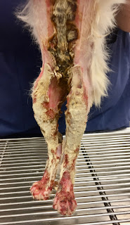
Maximus was non-ambulatory because the skin of his footpads was peeling off, leaving subcutaneous tissue exposed. Over the next several hours, the burned areas on his legs, chest, and belly gradually became purple in color. Some of the burned skin on his chest was becoming hard and leathery. (Fig. 1a,b) On a pain scale from 1-24 (1 being little or no pain, 24 being the worst pain possible), the attending clinician listed Maximus as 24. Intravenous lidocaine and ketamine were added to the fentanyl infusion to provide more analgesia. The dosage of fentanyl was also increased.

Fig. 1a: Maximus several hours after admission to the hospital

Fig. 1b


After sedating him with intravenous propofol, all of Maximus’ burned areas were carefully clipped and cleaned. With the hair removed, the burned regions were more clearly visible; the total surface area of his burned body appeared to be closer to 60%. 


More blood tests showed that Maximus’ albumin was already below normal. Albumin was being depleted because of the severe inflammation of the damaged skin and loss of albumin-rich fluid that oozed from his wounds. A nasogastric tube was placed to augment his dietary intake. 


Wound Management

On day two after admission, Manuka honey (Medihoney) was begun as the topical medication on the wounds, and standard bandaging was continued. The burned areas were gradually declaring themselves, and on day 4, some were starting to turn yellowish-brown and becoming hard and crusty. Escars were developing, indicating full-thickness skin injury. (Fig. 2)

Fig. 2: Escar development over mid abdomen and rear legs


To simplify the bandage changes that were time-consuming and painful, the attending clinician Dr. Becky Ball began covering Maximus with infant onesies, little pajama outfits that completely covered the entire body, legs, and feet. (Fig. 3) Each day the onesie was removed, the wounds gently cleaned, Medihoney ointment applied, and a clean onesie replaced. 

Fig. 3: Onesie pajama bandage applied.


Maximus was discharged 12 days after admission. The owner, a physician, assumed the nursing care at home. Rechecks with Dr. Ball were scheduled every 4-5 days for wound assessment. The escars gradually peeled away from the healthy skin, exposing new granulation tissue underneath. No evidence of infection was seen, and Maximus' appetite was good. 


Maximus continued to be re-examined about every two weeks. The eschars completely sloughed off, and the skin defects were contracting and closing well. But, as the skin tightened in his inguinal region, scar tissue pulled his rear legs medially, severely restricting his mobility. (Fig. 4) Surgery would be necessary to alleviate the contracture and allow Maximus to use the hind legs better. 

Fig. 4: Wound contraction of inguinal skin.

Skin Reconstruction


Seven months after Maximus’ injury, a caudal superficial epigastric axial pattern skin flap was performed to alleviate the inguinal wound contracture. (Fig. 5a-b) Click here for a detailed description of the skin flap procedure. The surgery went well, and he was discharged from the hospital the following day.

Fig. 5a: Axial pattern skin flap design. Arrow indicates the direction of rotation of the flap after it is dissected out.

Fig. 5b: Completed skin flap procedure.


Seven days postoperatively, suture removal was performed. The skin flap was viable and had healed well to adjacent skin. Maximus was much more mobile and less painful. Seven months postoperatively, he was examined again and was doing well with all wounds healed and his function back to normal. (Fig. 6, 7)

Fig. 6: Appearance of inguinal area seven months after skin flap reconstruction.

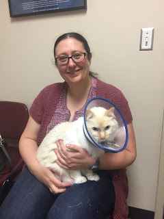
Fig. 7: Maximus and his owner 15 months after his injury.


Discussion


Maximus overcame a severe injury with burns over a large portion of his body. He exemplifies the remarkable wound healing process, particularly when supported with good nursing care. Manuka honey was effective in preventing infection and encouraging wound healing. Dietary support was instrumental in avoiding hypoproteinemia and malnutrition. Excessive wound contracture necessitated skin flap reconstruction, improving mobility and relieving chronic pain. 


Maximus' owner, a neonatologist, was instrumental in his recovery. She assumed the challenging nursing care at home and was diligent in the tedious work of medicating, bandaging, and maintaining good hygiene.


Maximus’ complete story is told in Dr. Birchard’s book: Their Tails Kept Wagging: Pets Show Us How Hope, Forgiveness, and Love Prevail, a collection of inspirational stories of severely ill pets who survived due to modern medicine and compassionate caregivers. For more information, click here.


Thursday, March 31, 2022

How Pets Can Help Heal the Trauma of Parental Estrangement


Their Tails Kept Wagging: Pets Show How Hope, Forgiveness, and Love Prevail
by Stephen J. Birchard
and Fe Anam Avis

Available on Amazon.com and other major booksellers' websites.

Tuesday, March 8, 2022

Their Tails Kept Wagging: Pets Show Us How Hope, Forgiveness, and Love Prevail


When seriously ill pets are surrounded with love, they are more likely to survive. 

In my book: Their Tails Kept Wagging: Pets Show Us How Hope, Forgiveness, and Love Prevail, we tell the stories of critically ill dogs and cats who 
refused to give up despite poor odds of survival. They lived because of modern medicine and the love and compassion of their caregivers and owners. 

For more information about the book, go to: Their Tails Kept Wagging


Stephen’s quotes


"For life is a seamless web. It connects us not merely with one another, but with all that is sentient; with all that shares its miracle of birth and feeling and death."— Abe Fortas

Tuesday, March 1, 2022

Reconstruction of Bilateral Caudal Maxillectomy Using an Autogenous Auricular Cartilage Graft

Stephen J. Birchard

Rebecca L. Ball


History and Physical Examination

Jack was a 4-year-old neutered male golden retriever that presented for inspiratory stertor for several weeks. (Fig. 1)

Fig. 1: Jack

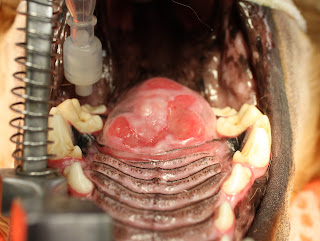
There was no other history of illness. On routine physical examination, no abnormalities were identified, but oral examination under sedation revealed a 4cm diameter firm circular mass in the caudal hard palate. (Fig. 2) The mass was not movable and extended the entire width of the hard palate between the molar teeth. 


Fig. 2: Caudal hard palate mass

Diagnostic Tests

Plain film radiographs of the thorax revealed no evidence of metastasis. CT imaging of the oral cavity showed invasion of the bone of the hard palate and extension of the mass into the nasal cavity. A punch biopsy of the mass was obtained, and histopathology revealed multilobular osteochondrosarcoma.


Surgical Treatment

Under general anesthesia, the mass was excised by bilateral caudal segmental maxillectomy, including excision of the molar teeth on each side. After incising the mucosa around the mass and dorsal to the teeth, the maxillary bones and hard palate were incised with a bone burr and osteotome. The mass penetrated the hard palate but was not invasive into the nasal turbinates. The mass was excised, and bleeding was controlled with electrocautery and Vetspon in the nasal cavity. (Fig. 3)

Fig. 3: Appearance of surgical site after resection of the mass.


The large oronasal defect was closed by constructing bilateral mucosal flaps using buccal mucosa on each side of the defect and then sliding them to the midline. The flaps were closed on the midline in 2 layers: 4-0 PDS simple interrupted in the submucosa, and 4-0 PDS simple interrupted in the mucosa and sutured to the mucosa of the hard palate and soft palate in a similar manner. (Fig. 4)


Fig. 4: Oronasal defect closed by bilateral buccal mucosal flaps.

The mass was submitted for histopathology. (Fig. 5) Multilobular osteosarcoma (MLO) was confirmed, and complete resection with clean margins was reported.


Fig. 5: Tissue specimen submitted for histopathology.

Postoperative Care and Complications

Five days postoperatively, Jack presented for anorexia and a foul odor from the mouth. Oral examination revealed partial dehiscence of the rostral and caudal aspects of the buccal mucosal flaps, creating oronasal fistulae at both sites. Conservative management was initially recommended to feed soft food meatballs followed by mouth flushing with clean water. 


Surgical Revision

The rostral oronasal fistula was repaired two weeks postoperatively using an autogenous auricular cartilage graft. Under general anesthesia, a 2 cm by 3 cm rectangular section of auricular cartilage was harvested from the inner leaf of the base of the left pinna. (Fig. 6) 

Fig. 6: Red rectangle indicates the approximate size and location of the harvested auricular cartilage graft. (This is not Jack but is a dog with similar size and anatomy of the ear pinna.)


After lightly debriding and undermining the mucosa along the perimeter of the oronasal defect, the cartilage was placed in the defect with the edges inserted underneath the mucosa. The cartilage was sutured to the mucosa with 4-0 PDS simple interrupted pattern. (Fig. 7) 

Fig. 7: Closure of mucosa over cartilage graft (yellow arrow) and remaining oronasal defect (green arrow) to be repaired later.


An esophagostomy tube was placed in Jack to allow feeding while bypassing the oral cavity.


Three weeks postoperatively, Jack presented to repair the remaining defect in his palate. The cranial defect had healed well over the cartilage graft. He was doing well with only occasional difficulty drinking water. Under general anesthesia, a hinged buccal mucosal flap was performed to close the caudal oronasal fistula. (Figs. 8)


Fig. 8: Closure of hinged buccal/pharyngeal mucosal flap over remaining defect. (Arrow indicates the direction of the inverted flap.)

Follow-up

Two weeks after this final reconstruction, Jack returned for a recheck examination. On oral exam, the flap was intact and healing well. (Fig. 9)

Fig. 9: Healed defect after flap revision. 


Three months later, another recheck examination showed complete healing of all flaps and no persistent oronasal fistulae. Jack was doing well clinically with normal eating, drinking, and activity levels. 


Jack lived 6 more years after his surgery to remove the MLO. He did well during that time with no issues related to his surgery and no tumor regrowth. He eventually died of causes unrelated to the MLO. Even with a prolonged recovery and multiple revision surgeries, the owners were pleased that they had pursued the treatment of Jack’s oral tumor. 


Discussion 

Bilateral segmental maxillectomy is a surgical option for oral neoplasia of the hard palate, mainly when the tumor is on the midline. Reconstruction is by creating bilateral sliding buccal mucosal flaps that are sutured to each other on the midline over the hard palate defect.

Dehiscence is common due to the tension across the incisions and the lack of underlying bone. Reconstruction of the resultant defect is challenging due to the scarcity of available local tissue. Autogenous auricular cartilage grafts have been reported to augment the closure of oronasal defects in dogs and cats. (1,2). This technique was effective in Jack repairing one of the defects in his mucosal closure. It provided a surface for the oral mucosa to proliferate and fill the oronasal defect. The remaining defect was judged to be too large to do an additional cartilage graft but healed with a hinged buccal/pharyngeal mucosal flap. The long-term results of the reconstruction surgeries were excellent. Further clinical studies are warrented to evaluate the auricular cartilage graft technique for other oral cavity disorders such as cleft palate.  


Acknowledgment

Dr. Becky Ball was the primary surgeon in all procedures performed on Jack and directed his postoperative care. 


References


1. Soukup JW, Snyder Cj, Gengler WR. Free Auricular Autograft for Repair of an Oronasal Fistula in a Dog. J Vet Dent, Summer; 26(2): 86-95, 2009


2. Cox CL, Hunt GB, Cadier MM. Repair of Oronasal Fistulae Using Auricular Cartilage Grafts in Five Cats. Vet Surg 36:164-169, 2007.


Friday, January 21, 2022

Traumatic Tracheal Avulsion in Cats: Diagnosis and Surgical Repair


Fig. 1: 6 mo. old DSH named Tigger


 Signalment and History

Tigger is a 6-month-old male neutered Domestic Short Hair cat who was presented for repair of a fractured left femur. (Fig. 1) Tigger had been hit by a car a few days before presentation. 

Physical Examination

On physical examination, the left rear leg was moderately swollen and crepitus was evident at the midshaft femur. The cat was also mildly dyspneic with tachypnea and mildly labored breathing. Thoracic auscultation was within normal limits. 

Diagnostic Evaluation

Thoracic radiographs showed pneumomediastinum and avulsion of the thoracic trachea with air-filled mediastinum seen bridging the gap. (Fig. 2) A mid-shaft oblique femoral fracture was also found. Tigger was transferred to the ICU for continuous monitoring and oxygen therapy if necessary. 

Fig. 2a: Lateral thoracic radiograph of Tigger showing the ruptured trachea 
and mediastinal "pseudo-airway." (white arrows)

                Fig. 2b: Ventro-dorsal thoracic radiograph of Tigger.
 

Anesthesia and Surgery

Tigger was sedated and an intravenous catheter was placed. The right side of his thorax was clipped and an initial cleansing of the surgical site was performed. Anesthesia was induced with propofol and an endotracheal carefully placed. The size of the endotracheal tube was smaller than indicated to avoid further injury to the trachea. Intermittent positive pressure ventilation (IPPV) was avoided to prevent disruption of the tenuous mediastinal connection between the ends of the ruptured trachea.

The right lateral thorax was prepared for aseptic surgery. A right lateral thoracotomy was performed at the 4thintercostal space. A Buford rib retractor was placed and the cranial thorax was explored. Dissection of the trachea confirmed a complete avulsion of the trachea midway between the thoracic inlet and the carina. The mediastinum was serving as the only connection between the 2 ends of the ruptured trachea. 

The mediastinum was carefully dissected and the proximal trachea exposed. The endotracheal tube was found within the tracheal lumen, grabbed with thumb forceps, and pulled farther out. The tube was then inserted into the distal tracheal segment. IPPV was then begun and maintained for the duration of the anesthetic episode. The ends of the ruptured trachea were healthy; no debridement was necessary. The tracheal anastomosis was performed with 4-0 PDS in a simple interrupted pattern. The sutures were placed by grabbing 1-2 tracheal rings on each side of the anastomosis and were full-thickness bites from outside to inside, exiting the tracheal lumen. (Fig. 3) All sutures were pre-placed to allow accurate placement and then tied. (Fig. 4)

Fig. 3a: Surgical exposure of the trachea in Tigger after preplacing sutures across the gap. Note endotracheal tube (ET) within the tracheal lumen. 

Fig. 3b: Appearance of the trachea after completing the tracheal anastomosis.

Before closing the thorax, the endotracheal tube was gently pulled back and forth within the trachea to confirm that no sutures had penetrated the tube. A thoracic drain tube was placed and the thoracotomy closed routinely. The fractured femur was then repaired. Tigger recovered uneventfully from anesthesia. 

Postoperative Care

Tigger was returned to the ICU and given intravenous fluids and analgesics for pain. He did well overnight with stable vital signs and eupneic breathing. One day postoperatively Tigger was doing well with no dyspnea, normal vital signs, and a good appetite. Minimal amounts of air or fluid had been recovered through the thoracic drain tube so it was removed. Tigger continued to do well and was discharged from the hospital on the 2nd day postoperatively with continued analgesic therapy and instructions to keep him indoors and restrict activity. 

Tigger returned for a recheck examination 1 month postoperatively. The referring veterinarian had removed skin sutures 2 weeks after surgery and the thoracic and leg incisions were well healed. On physical examination, Tigger was bright and alert with pink mucous membranes and normal auscultation of the heart and lungs. He was weight bearing on the left rear leg. Repeat thoracic radiographs were normal with only a slight indentation of the tracheal lumen at the site of repair. (Fig. 5) Radiographs of the left femur showed normal progression of healing at the fracture site. Tigger was discharged with instructions to continue rest and return in 4 weeks for repeat radiographs of the left femur to assess continued healing.


Discussion


Tracheal avulsion in cats is rare but has been reported. (1). It is likely the result of blunt trauma that causes the head and neck to be hyperextended which stretches the trachea leads to rupture. The site of rupture is usually the thoracic trachea midway between the thoracic inlet and the carina. The airway lumen is maintained by the mediastinum resulting in a so-called “pseudo airway”, allowing the cats to continue ventilating. In one study some cats presented up to 3 weeks after the traumatic episode. (1)

Cats with tracheal avulsion present for variable signs of dyspnea. Cats with delayed diagnosis may have trachea stenosis as fibrosis occurs at each end of the ruptured segments. Removal of the stenotic tracheal rings may be necessary to facilitate an adequate lumen at the anastomosis. (2)

The anesthetic protocol for these cats is designed around providing adequate oxygen flow and ventilation without causing iatrogenic disruption of the pseudo trachea. After induction, endotracheal intubation on Tigger was performed carefully without attempting to bridge the gap with the tube which could cause more injury. IPPV was avoided until the endotracheal tube was manipulated by the surgeon to provide a secure pathway for the delivery of oxygen. Immediately after thoracotomy, the first objective was to quickly pull the endotracheal tube from the proximal tracheal segment and place it in the distal segment to allow ventilation with oxygen and anesthetic gas. The trachea could then be carefully repaired. 

Based upon our experience with Tigger and a review of the reported cases, the prognosis for cats with tracheal avulsion is good after a successful repair. Most cats show adequate healing of the airway and can go on to live a normal life. 


References

1.     R N White, C A Burton. Surgical management of intrathoracic tracheal avulsion in cats: long-term results in 9 consecutive cases. Vet Surg, Sep-Oct 2000;29(5):430-5

2.     Fingland RB. Obstructive upper airway disorders. In: Saunders Manual of Small Animal Practice, 3d edition, Birchard SJ, Sherding RG, eds., Elsevier, 2006, pg. 1663-1664.