Author

Stephen J. Birchard DVM, MS, Diplomate ACVS

Sunday, September 29, 2013

5 Ways To Become a Faster Surgeon


“Time is trauma!”  How many times have you heard that from the anesthesiologist or anesthesia technician monitoring your surgical patient? How does it make you feel when people comment on the duration of your surgery; pressured, annoyed, even angry? All understandable reactions. But, there is a scientific basis for that cliché. The longer the tissues are exposed to the air the more likely to have complications such as excessive inflammation and infection.1 During my residency I was taught that for every hour the incision is open the rate of infection doubles.2 Studies on human hospitals have found the clinics with higher case loads tend to have fewer surgical complications than the slower ones.3

In the 19th century surgical speed was essential because surgery was done without anesthesia. Rapid surgery was necessary to decrease pain and mortality. Dr. Robert Liston was famous for being the fastest surgeon of his time. He could amputate a person’s leg in 2 and ½ minutes! But, occasionally his intent on being fast led to serious mishaps such as when he accidentally removed a man’s testicles while amputating his leg, and cutting off his assistant’s fingers during another lightening fast operation.

Thankfully in modern day surgery we have general anesthesia and other support systems to minimize morbidity and mortality and eliminate the need for 2 and ½ minute amputations. But prolonged surgery time still leads to increased postoperative problems for the patient and takes its toll on the surgeon. The longer the surgery takes the more likely you and your assistants will become stressed, tired, hungry, and have full urinary bladders. Tired surgeons become inefficient, less patient, and less meticulous. Decisions become more difficult and are made with less confidence. The surgeon loses focus and direction.

There is a also a stigma to being a slow surgeon. People, particularly non-surgeons, tend to judge surgical skill by how fast the surgeon is rather than by the hand skills or the ability to “think on your feet”. Its not always fair, but it happens.

Performing an operation swiftly is just one of many aspects of being a good surgeon and actually not the most important one. Speed in surgery is, although important, somewhat overrated. Patient evaluation, owner communication, and decision making before surgery are more important elements to good surgical practice. The decision making process involves a careful analysis of the patient, its owner, and the evidence based medicine that is relevant to your patient. The surgeon’s dedication to these principles trumps having hands that are a blur on the operating table.

Although being a speedy surgeon is not absolutely essential, the clinical evidence is clear that shorter operative times result in fewer postoperative complications. Here are 5 things you can do right now to reduce the time your surgical patients spend on the operating table:
  1. Have a plan. Base that plan on a thorough review of all aspects of the patient’s clinical presentation and status. Review the current literature for up to date information on the disorder and the surgical technique. Review anatomy and the steps involved in the procedure. Get advice from others before the surgery, not during. Discuss your plan with your support staff.
  2. Use good quality instruments and sutures. Have them ready in the operating room. Poor quality tools and materials lead to frustration, stress, and delays.
  3. Have an assistant surgeon. An assistant saves time by retracting, handing you instruments, cutting sutures, and keeping the instrument table organized. A lot of time is wasted searching for instruments when they are piled up on the table. Veterinary technicians love to scrub in and get close to the action! Let them do it!
  4. Eliminate distractions. Tell your staff not to bother you while you're in surgery. If a question can wait, save it for later. Limit the number of people in the operating room to reduce noise and commotion. Play soft, pleasant music to keep you relaxed and at ease.
  5. Don’t force it. You can’t will yourself to be faster. Allow it to happen naturally as you gain experience and repetition with procedures. Experience is a wonderful teacher; you learn how to handle tissues effectively and atraumatically. Your hands will start to flow through the surgery with little wasted motion and less tissue manipulation. Live in the moment and enjoy it.

A clean, organized instrument table makes surgery
more efficient and less frustrating.

 Conclusion

Surgical skill is more about what’s between your ears than in your hands. Practice your technical skills and always strive to have better hands, but be a “thinking” surgeon. Learn from your mistakes, keep a positive outlook, and believe in yourself. Do that, and you will find surgery to be a very satisfying part of your veterinary career, and your patients will appreciate your knowledge and skill by having uncomplicated postoperative recoveries.

References

1. Manilich E, Vogel JD, Kiran RP, Church JM, Seyidova-Khoshknabi D, Remzi FH. Key factors associated with postoperative complications in patients undergoing colorectal surgery. Dis Colon Rectum. 2013 Jan;56(1):64-71.
2. Cruse PJ, Foord R. The epidemiology of wound infection. A 10-year prospective study of 62,939 wounds. Surg Clin North Am. 1980;60(1):27-40.
3. Talya Salz, Robert S. Sandler. The Effect of Hospital and Surgeon Volume on Outcomes for Rectal Cancer Surgery Clinical Gastroenterology and Hepatology 2008 6:11, pg 1185–1193

Acknowledgement

Thank you to my friend and mentor, Dr. Ronald M. Bright, for teaching me to be a thinking surgeon.

Friday, September 27, 2013

Case Outcome: Patriot the pitbull with acute vomiting


Based on Patriot's acute onset of vomiting and diarrhea, a palpable hard abdominal mass, and the mixed density opaque object on radiographs (Fig. 1-2), exploratory laparotomy was recommended to the owner.


Fig. 1: Lateral abdominal radiograph with opaque density (arrow)
Fig. 2: Ventrodorsal radiograph with opaque density. (arrow)

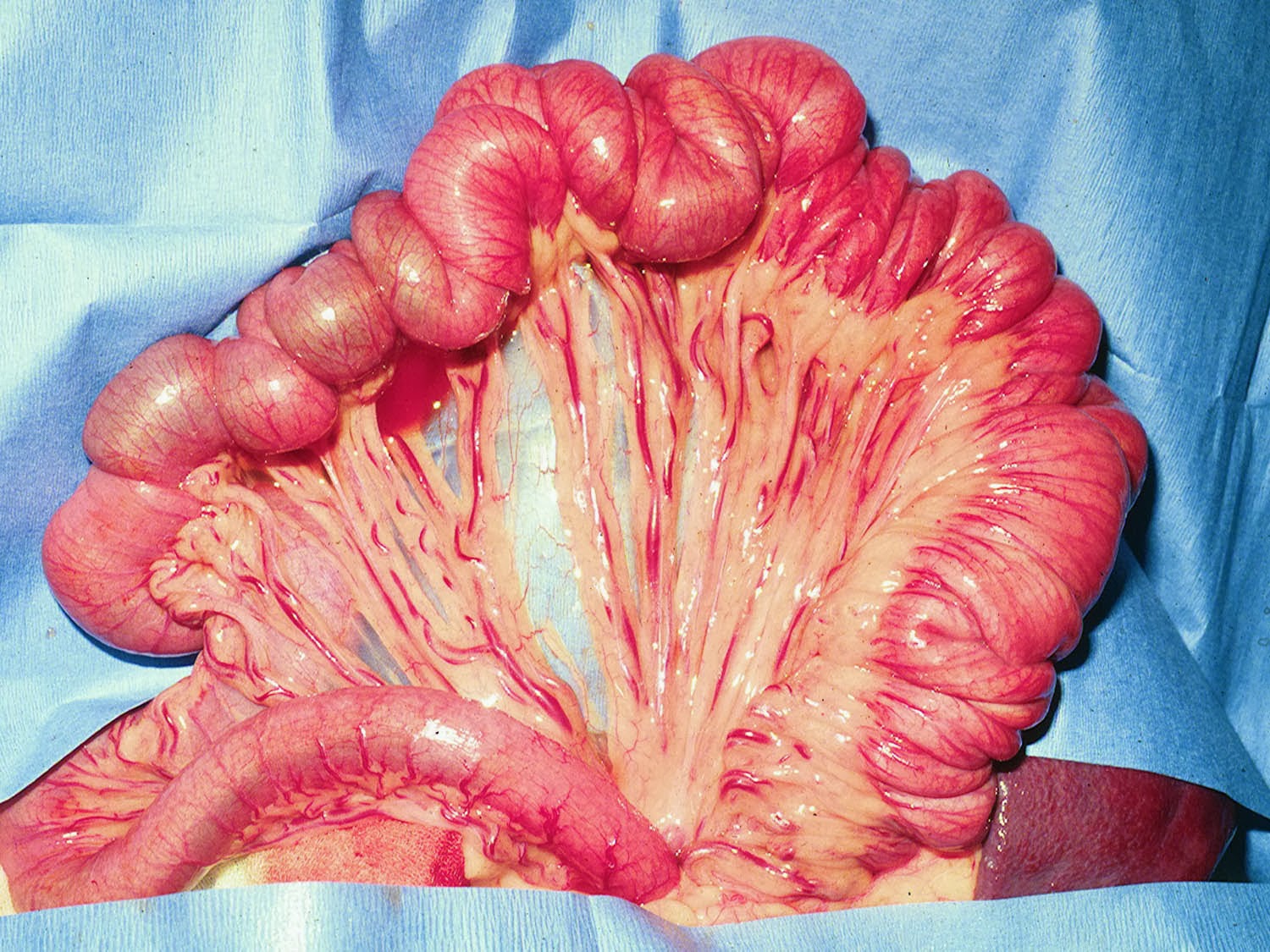
A ventral midline abdominal approach was performed. All structures were normal except for the ileo-ceco-colic area of the intestine. (Fig. 3-4)
Fig. 3: Ileo-ceco-colic area of intestine. The cecum was impacted with
firm material that was obstructing the ileum. (arrow)
The firm foreign body in the cecum and was manually massaged, fragmented, and moved into the colon. (Fig. 4) No incision in the cecum was necessary.
Fig. 4: Ileo-ceco-colic area after manually moving the foreign material into the colon.
Arrow indicates the now empty cecum.

Patriot made an uneventful recovery from surgery. He began eating and defecating normally several hours after surgery and was discharged 1 day postoperatively. One week after surgery the owner reported that he was doing well with no vomiting or diarrhea.

Comments

An interesting feature of this case was the severity of the vomiting even with a low (or distal) intestinal obstruction. The position of the foreign body on radiographs (cranial right abdomen) was consistent with a ileo-ceco-colic obstruction. The lack of intestinal dilation probably indicated a partial obstruction.  Ultrasonography or pneumocolon radiography would have been helpful to definitively localize the lesion.

Please post any questions or comments you have about this case either in the Comments box below or at: https://www.facebook.com/DrStephenBirchardVeterinaryCE

Thursday, September 26, 2013

What's Your Diagnosis and Treatment? Acute vomiting and diarrhea in a 3 year old pitbull.


Fig. 1: Patriot
Patriot is a 3-year-old male neutered 29 kg. pitbull that presented to Circle City Veterinary Specialty and Emergency Hospital with a history of acute vomiting and diarrhea.(Fig. 1) He had eaten bones 5 days ago and began vomiting 3 days prior to presentation.  He also had some episodes of diarrhea during this time.

On physical examination Patriot was bright and alert and slightly dehydrated. An egg sized firm movable mass was palpated in the mid-abdomen but otherwise his abdomen was soft and non-painful and no other abnormalities were found.

Complete blood count and serum chemistry profile were normal.

Lateral and ventrodorsal radiographs were obtained. (Figs. 2,3)
Fig. 2

Fig. 3

 What is your diagnosis?
 What is the next step for this patient?

Wednesday, September 25, 2013

Addendum to Linear Foreign Body Post; Look again at Fig 2a!

After reading yesterday's blog on linear foreign bodies, my friend Dr. Dave Biller (Head of Radiology at the College of Veterinary Medicine, Kansas State University) shared some valuable information and experience. At K State they routinely obtain left lateral abdominal radiographs for animals with a history of vomiting. This allows air to fill the pyloric antrum which may then outline a foreign body.

If you look closely at Fig 2a, you can see a foreign body in the pylorus. See the figure below with arrows pointing to the foreign body.


I honestly did not notice this until he pointed it out to me!

Yet another reason radiologists are such an important part of our profession. Thank you Dr. Biller!

Tuesday, September 24, 2013

Linear Foreign Bodies: One of the most sinister of all intestinal disorders


Fig. 1: String foreign body in the mouth of a cat

Linear intestinal foreign bodies can be serious and even life threatening. Besides causing intestinal inflammation and obstruction they can also cause multiple perforations at the mesenteric aspect of the bowel. Linear foreign bodies, such as string, fabric, and towels can involve only a few loops of bowel or they can extend from the stomach to the colon. If perforation has occurred the animal will develop septic peritonitis and rapidly deteriorate. Therefore surgery to remove linear intestinal foreign bodies should be considered urgent. In other words, “The sun should not set on a linear foreign body.”

Diagnosis

Animals with this type of foreign body usually present with acute vomiting and in some cases diarrhea. Lethargy and dehydration are common. Clinical signs are much more severe if septic peritonitis is present and can include signs of septic shock. Examination of the mouth in cats and less commonly dogs may reveal a string wrapped around the base of the tongue.(Fig. 1) Strings or other linear material may be seen protruding from the rectum. If a string is found around the tongue and also coming out of the rectum it is unwise to try to pull the string from either end.

Plain film abdominal radiographs show varying degrees of dilation and plication of the small intestine and in some cases bunching of the bowel in the abdomen. (Fig. 2-3) Gas pockets in the intestine that look like commas may be seen in the plicated areas. 
Fig. 2a: Lateral abdominal radiograph
of a dog with a linear intestinal foreign body.
Fig. 2b: Ventrodorsal radiograph of same dog in
Fig. 2a.
If a linear foreign body is suspected and an upper GI series is necessary to confirm the diagnosis, use a water-soluble contrast agent such as iohexol instead of barium because of the risk of leakage from intestinal perforations. Barium leakage into the peritoneal cavity worsens septic peritonitis by inhibiting phagocytosis of bacteria and causing a foreign body reaction. Ultrasound can be used to confirm the presence of the foreign body and plicated bowel and may also reveal peritoneal fluid that can be sampled and analyzed cytologically. Ultrasound has been shown to be superior to plain film radiography in diagnosing small intestine obstruction.1 Only 50% of dogs with linear foreign bodies showed intestinal dilation in that study.1
 
Fig. 3a: Lateral abdominal radiograph of a dog with a linear
foreign body. Less dilation of the bowel is seen compared to
dog in Fig. 2.

Fig. 3b: Ventrodorsal abdominal radiograph of same dog
as Fig. 3a. Note plicated duodenum (arrows)

Surgery

After a thorough abdominal exploratory, identify the affected areas of GI tract. (Fig. 4-5)
Fig. 4: Intestinal plication in same dog as Fig. 2 radiographs
Fig. 5: Intestinal plication in same dog as Fig. 3
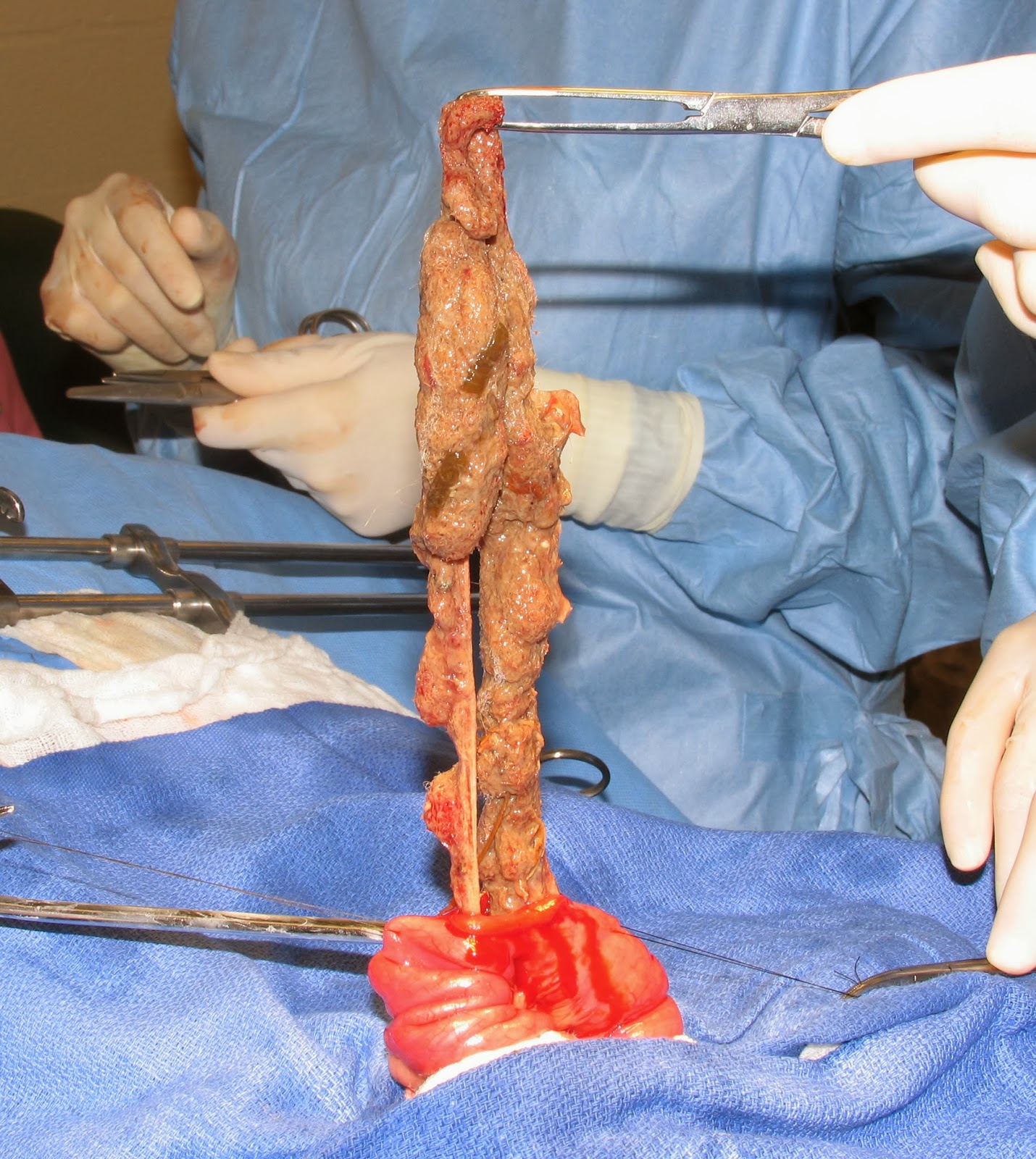
If no foreign body is present in the stomach make the enterotomy in the middle of the plicated bowel. I find that many times the entire linear foreign body can be removed through one enterotomy if you patiently apply gentle traction on the foreign body while gradually releasing the plication of the intestine with your other hand or with the assistant’s help.(Fig. 6) Use suction and abdominal sponges to prevent peritoneal contamination.

Fig. 6: Linear foreign body removed through one enterotomy.
String foreign bodies in cats are usually present in the stomach and intestine. If so, begin with a gastrotomy to remove that portion of the string. (See previous blog on gastrotomy) Gently pull the foreign body through the gastrotomy in attempt to remove the intestinal portion as well. If that is not possible remove as much as possible and then cut the string and close the gastrotomy. Remove the remainder of the string through one or more enterotomy incisions. (Fig. 7)
Fig. 7: String foreign body in a cat being removed through
an enterotomy
Also, prior to performing the enterotomy attempt to milk the foreign material into one segment of the intestine to make it easier to remove via a single enterotomy. Multiple incisions in the gastrointestinal tract were one risk factor for higher mortality in a large study of dogs and cats with intestinal foreign bodies.2 

Perform resection and anastomosis of bowel that has been perforated. Do not try to simply close the perforations; the tissue is not healthy and normal healing is unlikely. Try to avoid multiple anastomoses of the bowel; if possible include all the perforations in one resected segment so that only 1 anastomosis results.

If bowel perforation is present obtain samples of peritoneal fluid for culture, flush the abdomen with copious amounts of sterile saline, and place a closed suction drain (e.g. Jackson-Pratt drain).

Postoperative Care

See blog on intestinal biopsy for routine care of intestinal surgery patients. Postoperative care of animals with peritonitis will be covered in a future post. Animals with linear foreign bodies have a guarded prognosis compared to discrete, non-linear foreign bodies.

References

1. Sharma A, Thompson MS, Scrivani PV, et.al. Comparison of radiography and ultrasonography for diagnosing small-intestinal mechanical obstruction in vomiting dogs. Vet Radiol Ultrasound. 2011 May-Jun;52(3):248-55
2. Hayes G. Gastrointestinal foreign bodies in dogs and cats: a retrospective study of 208 cases. J Small Anim Pract. 2009 Nov;50(11):576-83.

Sunday, September 22, 2013

Blog Updates: NSAID Vote Final Tally, What's Coming Next Week, And I Have Questions For You

Final tally on NSAID preferences:


Which NSAID do you prefer for your canine patients?


Of course this is not a scientifically designed poll but Rimadyl was the clear choice amoung those who voted.

Don't forget to vote on which GI surgeries you feel comfortable doing. I like to ask questions like this because it helps me "gauge" my audience and influences what content I present in future posts.

More Discussion of the Blogs

I'm trying to figure out a way to get more discussion going on the blog website. I receive very few comments on the website itself, although many people comment on my Facebook page. Is that because you have to have a Google account to comment on the blog site? If you would rather not post a comment on the blog website, one option would be for you to put your comment or question on the facebook page:
https://www.facebook.com/DrStephenBirchardVeterinaryCE
Then I could copy and paste the comment or question and my answer on the blog for everyone to see. Of course I would make it anonymous in case you don't want your name on it. Another option would be to email your question to me (stephenbirchard@gmail.com) and then I could post it on the blog website.
Either of these options sound OK? I would love your input.

How Can We Reach More People?

I would also love your input on how we can have the blogs reach more vets, techs, and students. Not that I'm unhappy with the number of page visits we're getting but I'm just not confident that we always get the desired exposure on facebook that we want. How can we share the Vet Key Points website so that we can get more "traffic" as they say. (in general I hate "business-speak" like that but it seems like a good word here.) Do you belong to any online veterinary groups that might be interested in the site? Let me know any ideas you have.

What's Coming Next Week?

Look for more discussion of intestinal surgery topics, some "What's Your Diagnosis" cases, one that I did at Circle City Veterinary Specialty Hospital just last week, and  I'm working on making a video, using a model, of intestinal resection and anastomosis.

So, stay tuned!

Friday, September 20, 2013

STEVE! I Got a Bitch in the Truck!


His name was Ray. He was a middle-aged dog breeder; show dog handler, and boarding kennel owner.  He was a regular customer of the practice I worked in right out of veterinary school.  He was a big, tall, gruff man.  He was hard of hearing so he shouted all the time and you had to shout back. He was honest and direct and he expected the same from everyone else. His dogs were boxers; the females were “bitches” and the males were “studs”. Typical lingo in the dog-breeding world.

He always let himself in the back door of the practice. It was a courtesy granted him by the owner of the practice since he’d been a client for so many years. He never had an appointment; he didn’t need one. It was like he was a member of the staff. He came in the mornings before appointments started, usually on Mondays. He would come in the door and very loudly proclaim to my boss: “Doc, I got a bitch in the truck! I need you to look at her!” Then he would go back to the pick-up and fetch the dog.

He called me Steve, and at first he wasn’t so sure what to think of this “wet behind the ears” new graduate. My boss told him I had potential, but he had to find out for himself. He asked me to help hold a boxer while “Doc” did a rectal exam. Of course the dog started to squirm, and Ray yelled at me: “What’s the matter son, can’t restrain an animal? Didn’t they teach you that in vet school?” I was humiliated but held my tongue, and held the dog tighter. I found out later he was testing me. I guess I passed the test since I took his criticism in stride. The honest truth was, Ray knew more about dogs than I did and he and I both knew it. He intimidated me but I didn’t let him see it because I thought he wouldn’t respect me.

Ray had just enough veterinary knowledge to have a pretty good idea what the diagnosis and treatment was going to be when he brought a dog in. But, he didn’t always get the terminology quite right. If one of his dogs was lame in the rear leg, it had a rupture of the “crucial” ligament. If a puppy had a lump around the umbilicus, it had a “Biblical” hernia. I didn’t dare correct his mistakes. Ray didn’t know pathophysiology or pharmacology, but he knew dogs.
Gradually he started to trust me and let me treat his treasured boxers. He once asked me to remove a small skin mass from the head of one of his dogs. He wouldn’t let me give anything other than a local anesthetic, not even a sedative. He said: “Steve, if I tell her to stay, she won’t move a muscle.” I took the mass off with her sitting up and she didn’t budge.

We got to be friends and I developed a tremendous amount of respect for him. He even referred clients to me for veterinary care and I referred people to him for boarding.  After I moved on to Ohio State he occasionally called me for advice about his dogs. Underneath that crusty exterior was a heart of gold.  He loved his boxers more than anything. He taught me so much about dogs, things I never learned in vet school.  He also taught me about the world of dog breeding and show handling.  It's a tough way to scratch out a living, I can tell you that. He was an amazing man and I will never forget him.

My friend Ray is gone now.  I spoke with his daughter Pat today; she is 71 years young and told me that Ray passed away 7 years ago. The boxers have passed on, and the boarding kennel is closed. But Ray’s legacy and his spirit live on, and I can still hear that booming voice:

“STEVE! I got a bitch in the truck!”