Author

Stephen J. Birchard DVM, MS, Diplomate ACVS
Showing posts with label Oral cavity. Show all posts

Tuesday, March 1, 2022

Reconstruction of Bilateral Caudal Maxillectomy Using an Autogenous Auricular Cartilage Graft

Stephen J. Birchard

Rebecca L. Ball


History and Physical Examination

Jack was a 4-year-old neutered male golden retriever that presented for inspiratory stertor for several weeks. (Fig. 1)

Fig. 1: Jack

There was no other history of illness. On routine physical examination, no abnormalities were identified, but oral examination under sedation revealed a 4cm diameter firm circular mass in the caudal hard palate. (Fig. 2) The mass was not movable and extended the entire width of the hard palate between the molar teeth. 
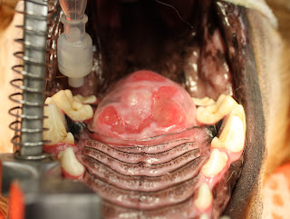

Fig. 2: Caudal hard palate mass

Diagnostic Tests

Plain film radiographs of the thorax revealed no evidence of metastasis. CT imaging of the oral cavity showed invasion of the bone of the hard palate and extension of the mass into the nasal cavity. A punch biopsy of the mass was obtained, and histopathology revealed multilobular osteochondrosarcoma.


Surgical Treatment

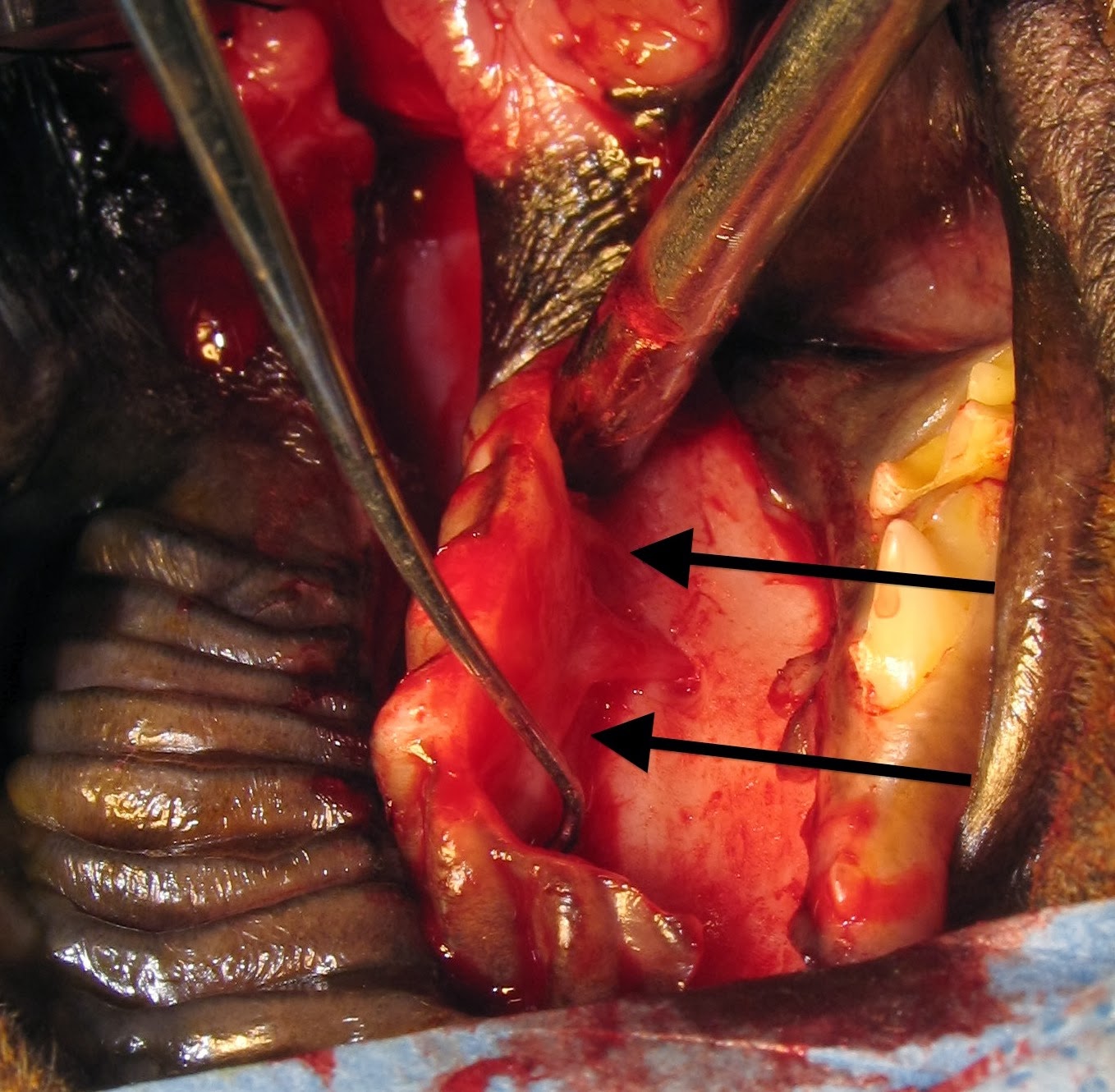
Under general anesthesia, the mass was excised by bilateral caudal segmental maxillectomy, including excision of the molar teeth on each side. After incising the mucosa around the mass and dorsal to the teeth, the maxillary bones and hard palate were incised with a bone burr and osteotome. The mass penetrated the hard palate but was not invasive into the nasal turbinates. The mass was excised, and bleeding was controlled with electrocautery and Vetspon in the nasal cavity. (Fig. 3)

Fig. 3: Appearance of surgical site after resection of the mass.


The large oronasal defect was closed by constructing bilateral mucosal flaps using buccal mucosa on each side of the defect and then sliding them to the midline. The flaps were closed on the midline in 2 layers: 4-0 PDS simple interrupted in the submucosa, and 4-0 PDS simple interrupted in the mucosa and sutured to the mucosa of the hard palate and soft palate in a similar manner. (Fig. 4)


Fig. 4: Oronasal defect closed by bilateral buccal mucosal flaps.

The mass was submitted for histopathology. (Fig. 5) Multilobular osteosarcoma (MLO) was confirmed, and complete resection with clean margins was reported.


Fig. 5: Tissue specimen submitted for histopathology.

Postoperative Care and Complications

Five days postoperatively, Jack presented for anorexia and a foul odor from the mouth. Oral examination revealed partial dehiscence of the rostral and caudal aspects of the buccal mucosal flaps, creating oronasal fistulae at both sites. Conservative management was initially recommended to feed soft food meatballs followed by mouth flushing with clean water. 


Surgical Revision

The rostral oronasal fistula was repaired two weeks postoperatively using an autogenous auricular cartilage graft. Under general anesthesia, a 2 cm by 3 cm rectangular section of auricular cartilage was harvested from the inner leaf of the base of the left pinna. (Fig. 6) 

Fig. 6: Red rectangle indicates the approximate size and location of the harvested auricular cartilage graft. (This is not Jack but is a dog with similar size and anatomy of the ear pinna.)


After lightly debriding and undermining the mucosa along the perimeter of the oronasal defect, the cartilage was placed in the defect with the edges inserted underneath the mucosa. The cartilage was sutured to the mucosa with 4-0 PDS simple interrupted pattern. (Fig. 7) 

Fig. 7: Closure of mucosa over cartilage graft (yellow arrow) and remaining oronasal defect (green arrow) to be repaired later.


An esophagostomy tube was placed in Jack to allow feeding while bypassing the oral cavity.


Three weeks postoperatively, Jack presented to repair the remaining defect in his palate. The cranial defect had healed well over the cartilage graft. He was doing well with only occasional difficulty drinking water. Under general anesthesia, a hinged buccal mucosal flap was performed to close the caudal oronasal fistula. (Figs. 8)


Fig. 8: Closure of hinged buccal/pharyngeal mucosal flap over remaining defect. (Arrow indicates the direction of the inverted flap.)

Follow-up

Two weeks after this final reconstruction, Jack returned for a recheck examination. On oral exam, the flap was intact and healing well. (Fig. 9)

Fig. 9: Healed defect after flap revision. 


Three months later, another recheck examination showed complete healing of all flaps and no persistent oronasal fistulae. Jack was doing well clinically with normal eating, drinking, and activity levels. 


Jack lived 6 more years after his surgery to remove the MLO. He did well during that time with no issues related to his surgery and no tumor regrowth. He eventually died of causes unrelated to the MLO. Even with a prolonged recovery and multiple revision surgeries, the owners were pleased that they had pursued the treatment of Jack’s oral tumor. 


Discussion 

Bilateral segmental maxillectomy is a surgical option for oral neoplasia of the hard palate, mainly when the tumor is on the midline. Reconstruction is by creating bilateral sliding buccal mucosal flaps that are sutured to each other on the midline over the hard palate defect.

Dehiscence is common due to the tension across the incisions and the lack of underlying bone. Reconstruction of the resultant defect is challenging due to the scarcity of available local tissue. Autogenous auricular cartilage grafts have been reported to augment the closure of oronasal defects in dogs and cats. (1,2). This technique was effective in Jack repairing one of the defects in his mucosal closure. It provided a surface for the oral mucosa to proliferate and fill the oronasal defect. The remaining defect was judged to be too large to do an additional cartilage graft but healed with a hinged buccal/pharyngeal mucosal flap. The long-term results of the reconstruction surgeries were excellent. Further clinical studies are warrented to evaluate the auricular cartilage graft technique for other oral cavity disorders such as cleft palate.  


Acknowledgment

Dr. Becky Ball was the primary surgeon in all procedures performed on Jack and directed his postoperative care. 


References


1. Soukup JW, Snyder Cj, Gengler WR. Free Auricular Autograft for Repair of an Oronasal Fistula in a Dog. J Vet Dent, Summer; 26(2): 86-95, 2009


2. Cox CL, Hunt GB, Cadier MM. Repair of Oronasal Fistulae Using Auricular Cartilage Grafts in Five Cats. Vet Surg 36:164-169, 2007.


Thursday, January 9, 2014

8 Principles of Cleft Palate Repair in Dogs and Cats

Fig. 1: Cleft hard palate (yellow arrow) and soft palate (red arrow) in a dog.
Congenital or acquired cleft palates are seen commonly in dogs and cats. (Fig. 1) Puppies and kittens should be checked for these defects immediately after birth. Early signs of cleft palate are regurgitation of food and water into the nose. Aspiration pneumonia can develop as a consequence. Clefts can involve the lips (primary) or hard and soft palate (secondary). Most clefts require surgical repair but dehiscence is common and some cases require multiple surgeries to achieve complete closure.

Principles of cleft palate repair:

Thoroughly evaluate the animal for rhinitis and aspiration pneumonia. Treat appropriately prior to surgery.

Preserve blood supply. The major palatine artery supplies the soft tissues of the hard palate and should be kept intact whenever possible.

Handle tissues gently. Use stay sutures on edges of mucosa and flaps to avoid excessive trauma.

Use fine, absorbable sutures. I prefer 4-0 polydioxanone since it has high tensile strength, is delayed absorbable, and is monofilament.

Avoid having sutures knots in the defect.

When making mucosal flaps to repair defects, make them as large as possible to avoid tension across the incision line.

Consider some kind of implant material when local tissues are not adequate for a tension free closure. (see future blog for an example)

Use an esophagostomy or gastrostomy tube postoperatively to bypass the mouth for nutrition to avoid food accumulation on the repaired palate.

Closure Technique

For most midline cleft palate the mucoperiosteal flap technique is an effective technique for closure. 
Fig. 2: Mucoperiosteal flap repair of a cleft hard palate in a dog.  A: dotted lines indicate incisions to make the
flap and elevate the mucosa on the opposite side; B: elevating the flap from the bone with a periosteal elevator; C: the flap has been inverted and sutured in place. Inset shows the horizontal mattress suture pattern from the edge of the flap to the overlying mucosa. (from: Marretta SM. Dentistry and diseases of the oropharynx. In: Saunders Manual of Small Animal Practice, editors Birchard and Sherding, 3rd edition, Elsevier, 2006, pg 609)

(Fig. 3) A large rectangular flap is made on one side of the cleft using a scalpel and periosteal elevator and is inverted. 
Fig. 3: The flap has been elevated and is being inverted to the other side (arrows).
This flap is tucked underneath the mucosa on the other side. Again using the scalpel and periosteal elevator, the mucoperiosteal tissues are elevated from the bone on the side opposite from the flap to allow the flap to be tucked into the space between the bone and the soft tissues. Use a horizontal mattress suture pattern to close the flap over the defect. (Figs. 2 and 4)
Fig. 4: the flap has been inverted from right to left, tucked under the mucosa  and sutured. The soft palate
cleft was closed by splitting the soft tissue layer on each side with a scalpel to separate the oral mucosal
side from the nasal mucosal side and sutured in 2 layers.
The denuded bone where the mucoperiosteal flap was harvested will become covered with granulation tissue and then mucosa in a few weeks. (Fig. 5)
Fig. 5: 2 weeks postoperatively after mucoperiosteal flap repair 

Postoperative Care

Postoperatively, as mentioned in the above principles, feed the animal through an esophagostomy or gastrostomy tube to avoid food accumulation on the incision.  Recheck the patient in 10-14 days to ensure progression of normal healing.

References

Howard DR, et.al. Mucoperiosteal flap technique for cleft palate repair in dogs.
J Am Vet Med Assoc. 1974 Aug 15;165(4):352-4.


,


Sunday, December 29, 2013

Salivary Mucoceles in Dogs: Cervical, Oral (rannula), and 1 type that causes airway obstruction

Anatomy


Fig. 1
(from: Manfra Marretta S. Dentistry and diseases of the oropharynx, in Birchard & Sherding (editors): Saunders Manual of Small Animal Practice, (3rd ed.). St. Louis. Elsevier, 2006, pp 632-635.)
The mandibular salivary gland is located at the bifurcation of the maxillary and linguofacial veins. (Fig. 1) The duct empties at the sublingual papilla, which is lateral to the most rostral aspect of the frenulum of the tongue. The gland has a well-defined capsule. The sublingual gland has two distinct portions, the monostomatic portion, which is immediately adjacent and rostral to the mandibular gland, and the polystomatic portion, which are small islands of gland dispersed along the salivary duct. The sublingual salivary duct is closely associated with the mandibular duct. The parotid gland is located at the base of the ear canal. Its duct empties adjacent to the upper 4th premolar. This is a wide, but thin gland; not globoid like the mandibular gland. The zygomatic gland is just ventral and medial to the zygomatic arch and the duct empties 1 cm caudal to the parotid duct.

Etiology of mucocele

 A salivary mucocele is the accumulation of saliva outside of the gland or duct system. The mucocele is not a true cyst since it does not have a secretory lining. It can be due to a variety of causes. Trauma to the head or neck that causes injury to either the gland or the duct can cause leakage of saliva. Oral mucocele (ranula) is a reported complication of mandibulectomy in dogs and cats. Infection or inflammation (sialadenitis) may cause enough tissue disruption to allow leakage. Histopathology of salivary tissue removed to treat mucoceles frequently reveals inflammatory changes. Although rare in dogs and cats but more common in humans, calculi can cause obstruction of the salivary duct with subsequent rupture of the duct and leakage. Rarely, neoplasia of the gland can cause disruption of glandular or ductal tissues.
Mucoceles are classified by their location. The cervical mucocele is the most common. A fluctuant mass is seen in the submandibular region and the leaking gland or duct is either the mandibular or the sublingual. If large, the mass may be mid-cervical or encompass the entire ventral aspect of the neck. An oral mucocele, also called a ranula, is due to leakage from the sublingual gland or duct and the swelling is intra-oral and lateral to the tongue. A pharyngeal mucocele is very similar to cervical but the fluctuant swelling is present in the pharynx, near the tonsil. Like the cervical mucocele, the pharyngeal is due to leakage from either the mandibular or sublingual salivary glands. Finally, the zygomatic mucocele can cause exophthalmus and/or peri-orbital swelling and is due to leakage from the zygomatic salivary gland.

Clinical Signs

Clinical signs of mucoceles vary according to their location.  The cervical mucocele causes a fluctuant, non-painful swelling in the cranial ventral cervical area. (Fig. 2)
Fig. 2: Cervical mucocele in a dog
 
The mass usually lateralizes to the affected side. However, in some cases the mass is directly on midline making it difficult to determine whether the right or left salivary glands are causing the problem. 
The oral mucocele, or rannula, can cause dysphagia, anorexia, excessive salivation, abnormal movements or protrusion of the tongue. (See previous post on Ollie for a case example.) The ranula may be large enough to be visible by the owner. It appears cystic and can be large enough to deviate the tongue to the opposite side.
            Dogs with a pharyngeal mucocele (Fig. 3) can present for inspiratory stertor and dyspnea since the fluid filled mass causes a physical obstruction in the pharynx and upper airway.
Fig. 3: Pharyngeal mucocele (arrow) in a poodle
Difficulty swallowing can also be a sign of pharyngeal mucocele.
A mucocele of the zygomatic salivary gland can cause exophthalmus, divergent strabismus, and a fluctuant swelling in the orbital area.

Diagnosis

            In most cases, salivary mucoceles are not a diagnostic challenge. The animals have a history of an acute or chronic fluctuant swelling that is not painful. Aspiration of the mass typically reveals a straw colored, mucinous fluid that appears ropey when pushed through a needle and syringe onto a slide. (Fig. 4)
Fig. 4: FNA of a mucocele (www.acvs.org)
Cytology of the fluid reveals few cells unless the mucocele is very chronic or infected in which case more evidence of inflammation is seen.
            Diagnostic imaging is usually not necessary but may be indicated in complicated cases or ones in which the presentation is atypical. Ultrasound or computed tomography may be helpful in animals with zygomatic mucoceles to differentiate the mass from neoplasia or foreign body.
            In some animals, the cervical mucocele is difficult to lateralize because it is either directly on midline or very large. Simple methods to determine the affected side are to lay the dog on its back and see to which side the mucocele gravitates. (Fig. 5)
Fig. 5: Placing the dog in dorsal recumbency can help lateralize the mucocele.
Sometimes by pushing medially on the mucocele while doing an oral exam the clinician can see one side of the pharyngeal wall bulge inward. In the rare case where the affected side is still unclear after these manipulations, positive contrast sialography can be performed. Contrast material is injected into the salivary duct and the resultant images studied for evidence of leakage or obstruction.

Treatment

            Regardless of location, the definitive treatment of a salivary mucocele involves removal of the offending salivary glands. Performing only drainage of the mucocele will not result in long-term resolution.
Surgical excision of mandibular and sublingual glands is the treatment of choice for cervical mucoceles. (Fig. 6)
Fig. 6: Excised mandibular (large gland to the left) and sublingual (remaining glands extending
from left to right) salvary glands
The mandibular gland, and the monostomatic and polystomatic portions of the sublingual gland are removed as a unit since their ducts are closely associated. The surgical approach is directly over the mandibular gland that lies just cranial to the bifurcation of the jugular vein into the maxillary and linguofacial veins. The capsule of the mandibular gland is incised to allow dissection and removal of the gland. Dissection then proceeds cranially along the mandibular duct. (Fig. 7)
Fig. 7: Surgical excision of mandibular and sublingual salivary glands in a dog.
Retraction of the digastricus muscle (arrow) facilitates dissection.
The multiple portions of sublingual gland are removed with the mandibular gland and the ducts. Dissect the ducts as far cranially as possible, then ligate and remove the tissues. Take care to avoid injury to the lingual nerve which lies over the salivary duct and serves as the most rostral limit of the gland and duct excision. Place a pen-rose or closed suction drain in the mucocele and close the incision routinely.
                        The etiology of pharyngeal mucoceles is similar to cervical mucoceles since they are also caused by leakage from the mandibular or sublingual glands. Therefore, remove these glands as described under cervical mucoceles. In addition, excise the pharyngeal mucocele via an intra-oral approach. Make an elliptical incision at the base of the mucocele, and excise the redundant mucosa and underlying tissues to be sure that the interior of the mucocele has been exposed. Although marsupialization of the resultant pharyngeal defect has been described, the author simply leaves this incision open to heal by second intention.
                        An oral mucocele, or ranula, is treated by “deroofing” the mucocele, followed by marsupialization. (See previous post on Ollie.) Deroofing is performed by simply removing the mucosa over the dorsal portion of the swelling being sure to expose the inside of the mucocele. Marsupialization is performed by suturing the inner lining of the mucocele to the oral mucosa. This allows the mucocele to remain open and drain into the mouth. Suturing is usually done with an absorbable suture such as Monocryl or PDS. Removal of the mandibular and sublingual salivary glands on the affected side is also be performed to prevent recurrence.
                        The zygomatic mucocele is treated by removal of the zygomatic salivary gland, which is located just ventral to the eye and medial to the zygomatic arch. Removal of the gland can be performed by partial removal of the zygomatic arch, or by ventral orbitotomy. The zygomatic arch can also be temporarily removed to provide exposure, and then reattached with orthopedic wire. Be careful not to injure orbital structures during the dissection.          

Postoperative Care

            Submit removed tissues for histopathology and culture. If a pen-rose drain was left in the mucocele, remove it once drainage becomes minimal. If infection was suspected or documented by positive cultures, treat the dog with appropriate antibiotics for at least 7-10 days. After marsupialization of a ranula, feed the dog soft food for 2-3 weeks, and flush the mouth with water or oral antiseptic lavage after eating.
            Complications after treatment of a salivary mucocele are rare. A seroma can occur due to the dead space created by the mucocele. Conservative treatment with warm compresses is usually effective. Recurrence of mucocele is rare but may indicate incomplete removal of the affected salivary tissue, or that the incorrect gland was removed.

References

Bellenger CR, Simpson DJ. Canine sialocoeles – 60 clinical cases. J of Sm. An. Practice 33:376-380, 1992.
Schmidt GM, Betts CW. Zygomatic salivary mucoceles in the dog. J Am Vet Med Assoc 172:940-942, 1978.
Knecht CD. Diseases of the salivary glands in the dog. Comp Cont Ed II:932-938, 1980
Bartoe JT, Brightman AH, Davidson HJ. Modified lateral orbitotomy for vision-sparing excision of a zygomatic mucocele in a dog. Vet Ophthalmology10:127-131, 2007.
Manfra Marretta S. Dentistry and diseases of the oropharynx, in Birchard & Sherding (editors): Saunders Manual of Small Animal Practice, (3rd ed.). St. Louis. Elsevier, 2006, pp 632-635.
Benjamino K, Birchard SJ, Niles JD, Penrod KD. Pharyngeal mucoceles in dogs: 14 cases. J Am Anim Hosp Assoc. 48(1):31-5, 2012

Tuesday, December 17, 2013

Outcome of Diagnosis and Treatment of "Ollie"

Fig. 1: FNA of the oral mass in Ollie

The gross appearance of the mass, its fluctuant nature, and the mucoid fluid that was aspirated (Fig. 1) were all suggestive of a rannula (oral mucocele). This is an accumulation of saliva from the sublingual salivary glands into the submucosal space in the mouth. 

The treatment for Ollie was marsupialization of the rannula (Fig. 2-3), and removal of the mandibular and sublingual salivary glands. (Fig. 4) 
Fig. 2: Completed marsupializaton of the rannula
Fig. 3: Close up of the marsupialized rannula showing the interior of the mucocele.

Marsupialization was performed by first incising over the dorsal aspect of the rannula with a scalpel. The saliva was evacuated from the cavity and the dorsal aspect of the rannula was debrided to make a large opening. The wall of the rannula was sutured to the inner tissue layer with 4-0 Monocryl in a simple continuous pattern. 

The right mandibular and sublingual salivary glands were removed in the standard fashion through a lateral cervical incision directly over the mandibular gland. (Fig. 4)
Fig. 4: Excised mandibular (yellow arrow) and sublingual (red arrows) salivary glands
in Ollie. Note the discolored areas of the sublingual glands. The salivary leakage was
likely originating from these glands.
Ollie was discharged the day after surgery with instructions to feed a soft diet and to flush the mouth with warm water after eating.

Ollie returned in 2 weeks for suture removal. The rannula had resolved and Ollie was eating and drinking normally and doing very well. 

In the next blog we will discuss salivary mucoceles in more detail.

Please post any questions you have about Ollie.

Monday, December 16, 2013

What's Your Diagnosis on "Ollie": A 5 year old Bichon with a huge oral mass?

Fig. 1
Ollie (Fig. 1) is a 5 year old male neutered Bichon who presented with a 5 day history of lethargy, discomfort, and excessive drooling. Oral exam revealed a large, soft mass just lateral to the frenulum of the tongue on the right side. (Fig. 2).
Fig. 2
Fine needle aspirate revealed a tan colored, thick, mucoid fluid. (Fig. 3)
Fig. 3

What is your diagnosis? What other diagnostic tests should be performed?
What is your recommended treatment?

Answer later this week!