Total ear canal ablation is removal of the entire ear canal. Indications for TECA include severe chronic otitis with obstruction of the canal, ear canal or middle ear neoplasia, severe trauma to the ear canal, or congenital abnormality that causes obstruction of the canal.
Key Point: Chronic proliferative otitis can result in calcification of the ear
canal. This is an irreversible, end stage change in the ear that can only be
resolved by TECA.
TECA is combined with a lateral bulla osteotomy (BO)
to remove residual epithelium and debris from the middle ear after the ear
canal is removed. Because of the prevalence of severe ear canal disease in dogs
and cats, TECA/BO has become a common surgical procedure. However, a properly
performed TECA/BO is a difficult procedure and can be associated with many
complications. It should be performed by a board certified surgeon who is
familiar with the anatomy of the ear and the technical aspects of the
procedure. However, a well performed TECA can significantly improve quality of
life of animals with ear disease.
Anatomy
The entrance of ear canal, the external acoustic
meatus, is surrounded by several cartilaginous structures including the tragus,
antitragus, helix, and antihelix. The external ear canal in dogs and cats is
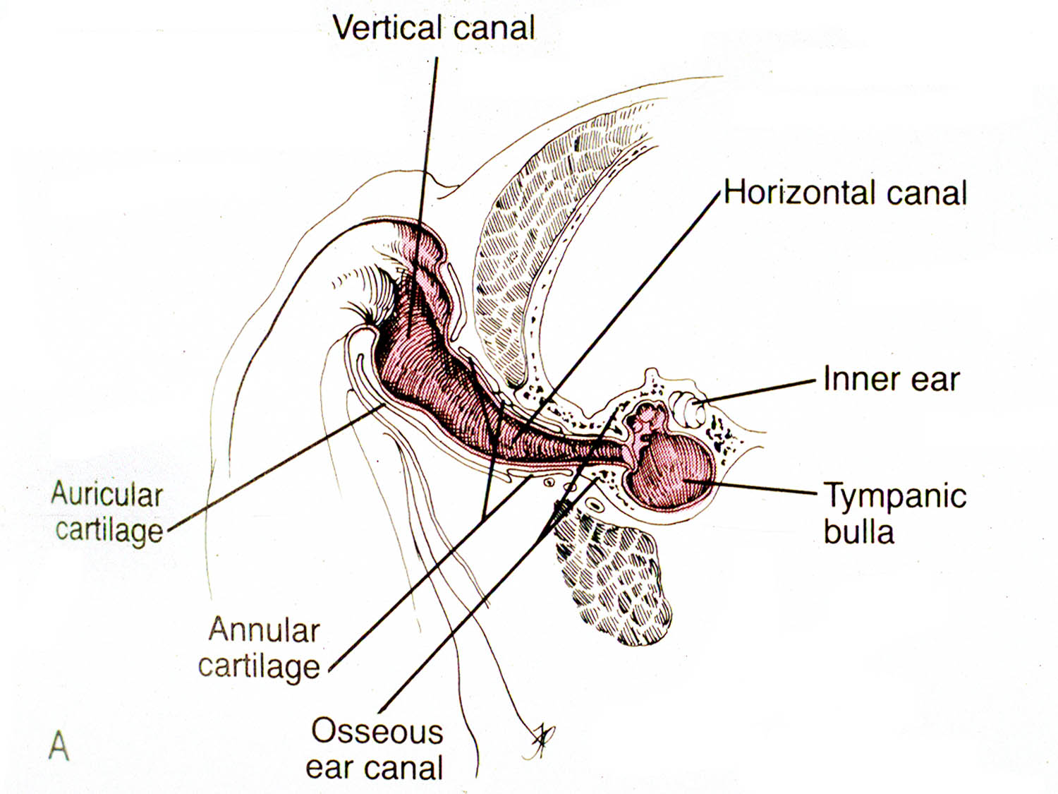
divided into vertical and horizontal portions. (Fig. 1)
 |
Fig 1: Cross section of the ear canal in a dog.
(from: Smeak DD. Surgery of the ear canal and pinna. Saunders Manual of Small Animal Practice, 3rd ed.,
Birchard and Sherding editors, Elsevier, 2006) |
The auricular cartilage
is the vertical portion of the canal. The annular cartilage is located where
the vertical canal turns into the horizontal canal. (Fig. 1)
The epidermal lining of the ear canal is rich in sebaceous
and apocrine glands. In dogs with chronic otitis externa, an epithelial pouch
may develop just adjacent and ventral to the tympanic bulla. (Fig. 2)
 |
Fig 2: Cross section of the ear in a dog with chronic otitis. Note the epithelial pouch
that develops in the canal adjacent to the entrance to the tympanic bulla. The tissue
colored red indicates that removed during a TECA/BO
(from: Smeak DD. Surgery of the ear canal and pinna. Saunders Manual of Small Animal Practice, 3rd ed.,
Birchard and Sherding editors, Elsevier, 2006) |
The
V-shaped parotid salivary gland lies over the ventro-lateral aspect of the
vertical ear canal.
The middle ear is located in the petrous temporal
bone. (Fig. 2) The tympanic bulla is the ventral wall of the tympanic cavity.
It is an air-filled cavity just medial to the tympanic membrane. In the cat a
septum divides the bulla into dorsomedial and ventrolateral compartments.
Sympathetic nerve fibers run through the middle ear, and adjacent to the tympanic
bulla are the facial nerve ventrolaterally, the carotid artery medially, and
the hypoglossal nerve ventrally.
The major blood supply to the ear is via the great
auricular artery and vein. Another important regional structure is the facial
nerve. The nerve exits the skull just caudal to the ear canal and courses
ventrally below the canal, then cranially. (Fig. 3)
 |
Fig. 3: Anatomy of important structures associated with the ear canal.
(from: Smeak DD. Surgery of the ear canal and pinna. Saunders Manual of Small Animal Practice, 3rd ed.,
Birchard and Sherding editors, Elsevier, 2006) |
The nerve is motor to the
lips and eyelids, therefore trauma to it causes lip droop and inability to
blink.
Preoperative
Considerations
Besides routine preoperative diagnostics such as
history, physical examination, and blood tests, a good otoscopic exam and
diagnostic imaging should be obtained on animals being considered for ear canal
ablation. Foreign bodies,
neoplasia, or obstructive disorders of the canal may be discovered on otoscopic
exam. Animals with tumors should also be screened for metastatic disease with
thoracic radiographs, fine needle aspirate of regional lymph nodes if enlarged,
and other tests as indicated. Skull radiographs or CT scan is usually
recommended before TECA/BO to assess the tympanic bulla. See Veterinary Key
Points blog “Nasopharyngeal Polyps in Cats”, 4/15/2015 for more discussion of
bulla imaging techniques.
Surgical
Procedure
After making the initial incisions around the
external acoustic meatus and then ventrally along the vertical canal, carefully
dissect the ear canal from surrounding tissues. (Fig. 4)
 |
Fig. 4: Initial dissection in a canine cadaver for a TECA/BO. (note: Figures 4,5,7, and 8 are cadaver specimens.
The cartilage around the external acoustic meatus has been severed
and lifted up to allow dissection down the canal. |
Dissect soft tissues
close to the canal to avoid trauma to important structures, such as the facial
nerve. (Fig. 5)
 |
| Fig. 5: Continued dissection of the canal exposing the facial nerve in the stay suture. (arrow) |
In ossified canals the facial nerve may be imbedded in the outer layer of the ear canal. (Fig. 6)
 |
Fig. 6: The facial nerve (surrounded by yellow vessel loops) is being released from its adherence
to the ossified ear canal (arrow). Note the groove left in the canal by the nerve after gently dissecting it off. |
After removal of the canal using scalpel or scissors, carefully
remove any remnants of canal and epithelium from the typanic bulla. Perform a
bulla osteotomy with rongeurs to better expose the interior of the bulla. (Fig. 7)
 |
Fig. 7: After removal of the canal, the tympanic bulla is exposed by removing some of the
lateral aspect of the bony wall with rongeurs. Any remnants of the ear canal attached to the bulla
can also be removed with the rongeurs |
Use a bone curette to remove epithelium and debris from the interior of the
tympanic bulla. (Fig. 8)
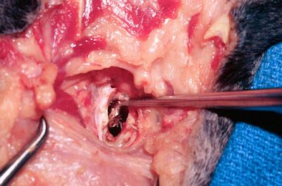 |
| Fig. 8: Curettage of the interior of the bulla with a bone curette. |
Avoid curettage of the dorsal aspect of the bulla to
prevent trauma to the structures of the inner ear. Submit samples of fluid or
debris from the tympanic bulla for culture and sensitivity. Also, submit the
ear canal for histopathology to rule out neoplasia. Flush the incision with
warm, sterile saline prior to closure. Close the incision in a “T” shape in
multiple layers: deep fascia, subcutaneous tissue, and skin.
Postoperative
Care and Complications
Postoperatively, protect the incision with a light
bandage or Elizabethan collar. Administer analgesics for at least 3-5 days
postoperatively. Long-term antibiotics (i.e. 3-4 weeks) are indicated in
animals with bacterial infections. Choose antibiotics based upon the results of
culture and sensitivity. If the animal’s eyelid motor function is decreased due
to facial nerve injury, keep the eye lubricated with eye ointments or drops (e.g.
Duratears) administered every 4-6 hours to prevent corneal ulcers until facial
nerve function returns.
Complications of TECA include acute pharyngeal edema, facial nerve damage, wound infection or dehiscence, Horner's syndrome, or deep abscesses. Deep abscesses occur due to leaving small amounts of secretory epithelium in or around the tympanic bulla. Reoperation to retrieve the residual epithelial tissue is usually necessary. Depending on the study, facial nerve deficits after TECA/BO in dogs can range from 36 to 48%, and in cats as high as 56%.(1-3) Although hearing is certainly diminished, some studies have found that some ability to hear is preserved even after removal of the ear canal.(2)
References
1. DD Smeak, WD DeHoff. Total Ear Canal Ablation Clinical Results in the Dog
and Cat. Veterinary
Surgery Volume 15, Issue 2, pages 161–170, March 1986
2. R. A.
S. White, C. J. Pomeroy. Total ear canal ablation
and lateral bulla osteotomy in the dog
Journal
of Small Animal Practice Volume 31, Issue 11, pages 547–553, November 1990
3. Rebecca
E. Spivack, A. Derrell Elkins, George E. Moore, and Gary C. Lantz (2013)
Postoperative Complications Following TECA-LBO in the Dog and Cat. Journal of the American Animal Hospital
Association: May/June 2013, Vol. 49, No. 3, pp. 160-168