Urethrostomy is the surgical procedure that creates a
permanent opening between the urethral lumen and the skin, and is indicated for
several disease conditions such as recurrent urinary obstruction due to
calculi, trauma or neoplasia.
Urethrostomy in the dog can also be performed when penile amputation is
necessary for neoplasia or other conditions, such as hypospadias.
Several types of
urethrostomy have been described for dogs, but scrotal urethrostomy is
currently the procedure of choice.(1-3) After castration and scrotal ablation,
the urethra is opened at the level of the scrotum and sutured to the adjacent
skin.
Advantages of scrotal urethrostomy are:
- the urethrostomy stoma is located ventrally on the dog, minimizing urine scald of surrounding skin
- the urethra is relatively close to the skin at this location
- the scrotal portion of urethra is large enough to allow formation of a large stoma that allows passage of calculi and rarely strictures
- urinary continence is not compromised by developing a stoma at this location.
Surgical Technique for Scrotal
Urethrostomy
Under general
anesthesia place the dog in ventral recumbency. If possible, pass a urinary
catheter to flush calculi retrograde into the urinary bladder (if applicable),
and leave in to help with identification of the urethra during dissection.
Prepare the scrotum, ventral abdomen, and ventral perineum for aseptic surgery.
Make an elliptical incision around the base of the scrotum. Be sure to leave sufficient skin to allow for a tension-free closure of skin to urethral mucosa. Perform scrotal ablation and castration in a routine fashion. (Fig. 1)
 |
| Fig. 1: Beginning of a scrotal ablation by performing castration and scrotal ablation. |
Dissect through the subcutaneous tissue and identify the retractor penis
muscle. (Fig. 2) Dissect the muscle away from the urethra and retract it laterally
to the urethra using forceps or a stay suture.
 |
| Fig. 2: Dissection through the subcutaneous tissue exposing the retractor penis muscle (black arrow), and urethra(white arrow). |
Incise the
urethra on the midline with a scalpel (4-6 cm opening). (Fig. 3a,b)
 |
| Fig. 3a: Incision is made on the urethral midline with a scalpel. The incision can be started with a scalpel and extended with fine scissors. |
 |
| Fig. 3b: Completed urethral incision. Note urinary catheter in the urethral lumen. |
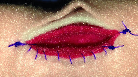
Suture the urethral mucosa and submucosa to the skin with fine suture material (4-0 PDS or Monocryl, swaged-on taper needle) in a simple continuous pattern. (Figs. 4-9) Start the suture line at the caudal aspect of the incision, and work cranially.
 |
| Fig. 4: Surgical model of the penis and tissue layers important for urethrostomy. (TA = tunica albuginea) |
 |
| Fig. 5: Closure of the urethrostomy begins with 2 lines of sutures at the caudal aspect of the incision. |
 |
| Fig. 7: Completion of one side of the closure. |
 |
| Fig. 8: Completion of both sides of the urethrostomy |
Avoid excessive
manipulation of the urethral mucosa since it is friable and will bleed more
with trauma.
 |
| Fig. 9: Appearance of a healed scrotal urethrostomy 2 weeks postoperatively. |
Postoperative Care
Typical
postoperative care after scrotal urethrostomy involves prevention of incisional
trauma by using an Elizabethan collar on the dog, applying petroleum jelly
around the incision to keep it moist and clean, close monitoring of the
incision for swelling or bruising, and general supportive care (e.g., analgesics).
Light sedation with acepromazine (0.05 mg/kg, SQ or IM) can be helpful to reduce
hemorrhage from the incision site which is the most common postoperative complication. Suture removal is not necessary when
absorbable suture is used to close the urethrostomy. If persistent hemorrhage
occurs (i.e., for several days after surgery), carefully re-assess the incision
for areas where the mucosa has not properly healed to the skin. Additional sutures in these areas to
close the defect should alleviate the problem.
Postoperative
urethral stricture, although a possible complication of urethral surgery, is
uncommon in a well-performed scrotal urethrostomy. Stricture may occur due to
chronic licking of the incision or poor apposition of the urethral mucosa to skin
during closure. Treatment of stricture is to revise the urethrostomy and insure
meticulous mucosa to skin closure.
References
1. Bilbrey SA,
Birchard SJ, Smeak DD. Scrotal urethrostomy: A retrospective review of 38 dogs
(1973 through 1988). J Am An Hosp Assoc 1991; 27: 560-564.
2. Newton JD,
Smeak DD. Simple continuous closure of canine scrotal urethrostomy: Results in
20 cases. J Am An Hosp Assoc 1996; 32:531-534.
3. Collins RL,
Birchard SJ, Chew DJ, Heuter KJ. Surgical treatment of urate calculi in
Dalmatians: 38 cases (1980-1995). J Am Vet Med Assoc 1998; 213:833-838.


No comments:
Post a Comment