Author

Stephen J. Birchard DVM, MS, Diplomate ACVS

Tuesday, November 18, 2014

Intravenous Fluids in Anesthetized Dogs and Cats: Are we giving too much?

Intravenous fluid therapy is one of the most important perioperative treatments veterinarians provide for their patients. Intravenous fluids are considered a necessary part of the anesthesia protocol because of hypotension and vasodilation that can occur due to the anesthetic drugs. 

All animals being prepared for anesthesia and surgery need to be assessed for hydration status and disorders that create fluid losses, e.g. vomiting and diarrhea. Intravenous fluid dosages will be influenced by the animal’s current hydration and ongoing fluid losses. Intravenous fluid dosages may also be affected by disorders that could predispose the animal to over-hydration such as cardiac or renal disease.

The traditional intravenous fluid rate for healthy animals under anesthesia has been 10ml/kg/hour.(1) In the recent AAHA/AAFP fluid therapy guidelines, this recommendation has been revised.(2) Table 4 from the paper describes current fluid therapy guidelines for anesthetized cats and dogs:

Table: Recommendations for Anesthetic Fluid Rates (from: 2013 AAHA/AAFP Fluid Therapy Guidelines for Dogs and Cats. Harold Davis, BA, RVT, VTS (ECC), Tracey Jensen, DVM, DABVP, Anthony Johnson, DVM, DACVECC, et.al., J Am Anim Hosp Assoc 2013; 49:149–159)

- Provide the maintenance rate plus any necessary replacement rate at <10 mL/kg/hr
- Adjust amount and type of fluids based on patient assessment and monitoring
- The rate is lower in cats than in dogs, and lower in patients with cardiovascular and renal disease
- Reduce fluid administration rate if anesthetic procedure lasts 1 hr
- A typical guideline would be to reduce the anesthetic fluid rate by 25% q hr
until maintenance rates are reached, provided the patient remains stable

Rule of thumb for cats for initial rate: 3 mL/kg/hr
Rule of thumb for dogs for initial rate: 5 mL/kg/hr

Note that not only are the initial fluid rates lower than the previously recommended 10ml/kg/hr, but a schedule for gradual reduction of fluid rates as the anesthetic period progresses is also recommended. These guidelines are considerably different from what was previously thought to be necessary fluid rates for anesthetized animals, but are based on carefully considered factors, evidence based medicine, and clinical experience of board certified specialists.

References

1. Ann Weil, DVM, DACVAA. Anesthesia reboot: Erase these myths and misconceptions. Veterinary Medicine, October 2014, pg. 318.

2. Harold Davis, BA, RVT, VTS (ECC), Tracey Jensen, DVM, DABVP, Anthony Johnson, DVM, DACVECC, et.al. 2013 AAHA/AAFP Fluid Therapy Guidelines for Dogs and Cats. J Am Anim Hosp Assoc 2013; 49:149–159.

Questions:

What are your thoughts or opinions about this change in recommended fluid dosage?

In private practice, in which anesthetized patients do you typically run intravenous fluids; in all animals or do you have some kind of selection criteria? In other words, what do you think the standard of care should be for fluid administration under anesthesia?

Please post comments either here on the blog site or on my facebook page:
Dr. Stephen Birchard, Veterinary Continuing Education

.

Monday, November 10, 2014

Large Tumor On The Skull of a Dog: The story of "Rocky" the Norwegian Elkhound

Rocky was a 7 year old male castrated Norwegian Elkhound. Several months previous to presentation he developed a slowly growing firm mass on the dorsal cranium.  The owners reported no other significant health problems. 
Fig. 1: This is Rocky under anesthesia being prepared for surgery.
Note large mass on the dorsum of the skull.
On physical examination the mass was well circumscribed but not movable.(Fig. 1) Neurologic exam revealed mild posterior paresis. In all other respects Rocky was behaving like a normal dog.

Diagnostic Evaluation

Radiographic evaluation found no evidence of metastasis of the mass in the lungs or abdomen.  A complete blood count and serum chemistry profile was within normal limits. MRI of the skull showed a very large ossified mass of the dorsal cranium extending ventrally into the cranial vault.(Fig. 2)
Fig. 2: MRI of Rocky's head. Note large skull mass (arrows) that extends ventrally.
Surgery

Although Rocky’s owners were warned about risks of surgery such as brain injury, hemorrhage, infection, and possible incomplete resection of the mass, they wanted to pursue surgical removal. They also knew that the most likely neoplasms causing the tumor were osteosarcoma or multilobular osteochrondrosarcoma.

Rocky was anesthetized and placed in sternal recumbency. His entire head was prepared for aseptic surgery. (Fig. 3)
Fig. 3: Preoperative view of Rocky just prior to beginning of surgery
A midline approach over the mass was performed. Sharply dissecting soft tissues including the temporalis muscles, and retracting them laterally, easily exposed the mass. (Fig. 4) 
Fig. 4: Surgical exposure of the skull mass after dissection of the soft tissues.
Dorsal is to the top of the photograph.
Careful blunt and sharp dissection at the base of the mass allowed for gradual moblilization, and the mass and the involved cranium then easily lifted off the remainder of the skull and were excised. (Figs. 5,6)
Fig. 5: Most of the skull mass has been reflected off the remainder of the skull and is
being held upside down in my hand.
Fig. 6: The resulting skull defect after complete removal of the mass.
Moderate hemorrhage occurred from remnants of tumor attached to the dura and at the edges of the skull. Hemostasis was achieved with judicious electrocautery and hemostatic sponges. Careful debridement of the remaining gross tumor was attempted but was difficult because of poor exposure due to recurrent hemorrhage, and attachment of the mass to the dura.

The skull defect was filled with an autogenous fat graft that was aseptically harvested from Rocky’s lumbar area. The temporalis muscles were then mobilized by undermining them from the skull and used as the first layer of closure by suturing them together on the midline. The remaining tissue layers were closed routinely.(Fig. 7)
Fig. 7: Closure of the temporalis muscles over the skull defect
Postoperative Care and Follow Up

Rocky made an uneventful recovery from anesthesia. Repeat neurologic examination revealed no change from his preoperative status. Routine supportive care and analgesic therapy was administered.

Rocky was discharged from the hospital several days postoperatively. Histopathology of the mass revealed a multilobular osteochondrosarcoma. Two weeks later he was examined for suture removal and was doing very well, including improvement of his posterior paresis. He was rechecked again several weeks later and was continuing to do well.(Fig. 8)
Fig. 8: Rocky at his recheck examination several weeks postoperatively
The owners declined any adjunctive therapy such as radiation or chemotherapy, for the tumor.  Approximately 9 months postoperatively Rocky’s mass began to re-grow. Several months later the owners elected to have Rocky euthanized. 

Discussion

Multilobular osteochondrosarcoma (MLO) is a well-described tumor of bone and frequently found on the skull in dogs.(1) The tumor is typically slow growing but locally invasive and can be metastatic. Complete resection of a small MLO can have a good prognosis. However, recurrence of tumor commonly occurs after incomplete resection (approximately 50% of cases) as was the case in Rocky. After surgical treatment, disease free interval of 288-1332 days has been found depending on tumor grade. (2)

Options for reconstruction of large defects of the cranium include rigid materials like polymethylmethacrylate, mesh implants, or autogenous soft tissues using the temporalis muscles. (3) An autogenous fat graft to protect the dura combined with temporalis muscle reconstruction was an effective option for Rocky.

An extraordinary aspect of this case is that Rocky had severe compression of his cerebrum by the tumor, yet had only mild neurologic signs. He also made a surprisingly uneventful recovery from surgery and lived about 1 year postoperatively. Of course we would have preferred to achieve a complete resection of the mass but that was not possible without risking serious complications.

Post questions or comments about Rocky either here or on my Facebook page, Dr. Stephen Birchard, Veterinary Continuing Education. Thanks!

References

Multilobular osteochondrosarcoma of the canine skull: 16 cases (1978-1988).
JAAHA 1989, 195(12):1764-1769]

2. Veterinary Society of Surgical Oncology website: http://www.vsso.org/Bone_MLO.html


3. Boston, SE. Craniectomy and orbitectomy in dogs and cats. Can Vet J. May 2010; 51(5): 537–540.

Monday, November 3, 2014

Closure of Elliptical Incisions in Dogs: The "Rule of Halves"

Elliptical incisions commonly result from removal of skin tumors, other lesions, or debridement of traumatic wounds. Since loss of a section of skin occurs, the closure of the defect can result in so called “dog-ears” at each end of the incision (small flaps of skin that protrude from the ends of the incision).  An easy method to prevent dog-ears and create a cosmetic and secure closure is the “rule of halves” technique, also called bisectional closure. (Fig. 1)
Fig. 1: The rule of halves for closing elliptical incisions.
Dotted lines represent placement of sutures
This technique is used for the deep fascia and subcutaneous layers of the wound. The sutures below the skin relieve the tension across the incision and align the skin edges making the final suturing of the skin much easier.

Technique for the Rule of Halves Closure

Take the first suture bite of the deep fascia and/or subcutaneous tissue in the middle of the incision. (Fig. 2)
Fig. 2: Surgical model of elliptical incision.
The subcutaneous tissue is red, the skin is pink.
Dotted line indicates where first subcutaneous suture is placed.
This divides the incision into 2 equal parts. (Fig. 3)
Fig. 3: First subcutaneous suture has been placed.
Dotted lines indicate placement of the next 2 sutures.
Now take suture bites in the middle of each of the 2 defects, then in the middle of the 4 defects, and so on until the subcutaneous layer is completely closed. (Fig. 4) 
Fig. 4: Final subcutaneous suture being placed. Note that the suture knot
is buried by taking the first bite, from inside out, on the side closest to the surgeon.
Then suture the skin routinely. (Fig. 5)
 
Fig 5: Completed closure

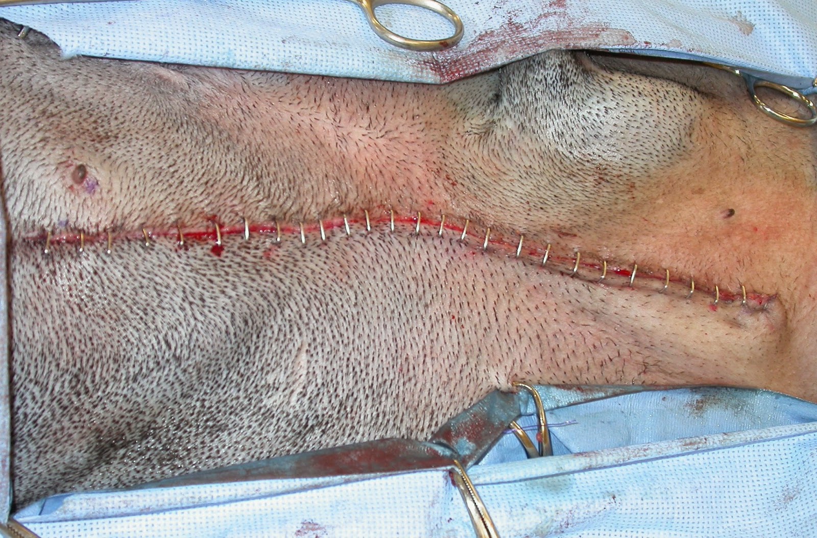
This technique for wound suturing, although somewhat more time consuming than doing simple continuous patterns for the subcutaneous layer, results in a very secure and cosmetic closure. (Figs. 6-8)
Fig. 6: Proposed lines for excision of a mast cell tumor in a dog.  (X = tumor,
circle indicates 3 cm margins, elliptical lines are proposed incision)
Fig. 7: Same dog as in Fig. 6 after excision of the tumor and surrounding skin.
Dotted line indicates location of first suture.
Fig. 8: Completed closure of same dog in Fig. 6.

Post questions or comments here or on my Facebook page (Dr. Stephen Birchard, Veterinary Continuing Education)