Author

Stephen J. Birchard DVM, MS, Diplomate ACVS

Monday, May 19, 2025

The Punch Technique for Aural (Ear) Hematomas in Dogs and Cats: Simple, effective, and cosmetic!


Auricular hematomas occur from hemorrhage that develops between the 2 leaves of cartilage of the pinna. The hemorrhage can occur due to trauma to the pinna from head shaking or scratching.  Inflammatory conditions of the ear canal, such as ear mites, foreign bodies, or bacterial otitis may be the inciting cause.

Diagnosis

Aural hematomas are characteristic in appearance.(Fig.1,2) 

Fig. 1: Aural hematoma in a cat
Fig. 2: Aural hematoma in a young white tiger

The pinna is enlarged and fluctuant. Differential diagnoses include acute allergic response causing severe swelling of the pinna, and neoplasia of the pinna. Aspiration of the mass reveals blood or serum. Rule out underlying ear canal problems by a thorough palpation of the ear canal and otoscopic exam. Also, thoroughly examine the animal for evidence of skin disease such as allergies, seborrhea, fleas, or pyoderma. 


Treatment

Many methods have been described for treatment of aural hematomas. Incision and drainage, drain tubes, and laser techniques have all been described.(1-3) Medical management by simple drainage combined with either systemic or local corticosteroid therapy has also been advocated. The advantage of medical therapy or simple needle drainage is excellent cosmetic result. However, incidence of recurrence with these treatments is high. The advantage of incision and suture is a low rate of recurrence, but the scaring of the pinna can cause poor cosmetic results.

The punch technique described here (Fig. 3) allows effective drainage and very low incidence of recurrence.(4) The cosmetic results are also very good since little scar tissue develops in the small incisions.
Fig. 3: Depiction of punch technique for aural hematomas in dogs and cats.
(from: Smeak DD. Surgery of the ear canal and pinna. Saunders Manual of Small Animal Practice, 3rd ed.,
Birchard and Sherding editors, Figure 60-1, Elsevier, 2006, pg. 583)

Surgical Technique
  • Clip and prepare both sides of the pinna for aseptic surgery. Place a surgical sponge in the ear canal to prevent accumulation of blood.
  • Use a skin biopsy punch (size 4-6 depending on the size of the dog) to remove small plugs of skin and cartilage on the medial side of the pinna.(Fig. 4) 
    Creating punch incisions on the medial aspect of the pinna for  drainage of aural hematoma.
  • Attempt to penetrate only the skin and 1 layer of the cartilage with the punch; however inadvertent removal a small section of both of the cartilage layers is not problematic.
  • Make incisions about 0.5 – 1 cm apart and perform as many punches as necessary to drain the entire hematoma. 
  • Tack the skin edge of each incision with monofilament nylon, polypropylene, or Monocryl in a simple interrupted pattern.(Fig. 5) The size of suture can be 3-0 or 4-0 depending on the size of the animal. It is not necessary for the suture to penetrate full thickness through all layers of the pinna including the skin on both sides but the suture should incorporate both layers of cartilage and the skin on the medial surface.

Fig. 5: Suturing the edge of each punch incision with monofilament suture.

Postoperative Care

Postoperatively, place a stockinette on the dog’s head to protect the pinna and reduce bleeding. I prefer not to send dogs home with a full bandage on the ear or head. Keep the dog from scratching the ear with an Elizabethan collar. Remove sutures at 14 days.(Fig. 6)
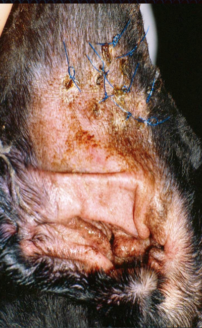
Fig. 6: Pinna of a dog 2 weeks after the punch technique
for aural hematoma (Photo courtesy of Dr. Daniel Smeak)
If otitis externa or other skin disorder is present, treat appropriately.

References

1. Pavletic MM Use of laterally placed vacuum drains for management of aural hematomas in five dogs. J Am Vet Med Assoc. 2015 Jan 1;246(1):112-7.
2. Dye TL, Teague HD, Ostwald DA Jr, Ferreira SD. Evaluation of a technique using the carbon dioxide laser for the treatment of aural hematomas. J Am Anim Hosp Assoc. 2002 Jul-Aug;38(4):385-90.
3. Kagan KG Treatment of canine aural hematoma with an indwelling drain. J Am Vet Med Assoc. 1983 Nov 1;183(9):972-
4. Smeak DD. Surgery of the ear canal and pinna. Saunders Manual of Small Animal Practice, 3rd ed., Birchard and Sherding editors, Elsevier, 2006, pg. 582)

Blog Update: Dr. Birchard has published a new book: "Their Tails Kept Wagging", a collection of moving stories about pets with serious illness who survived. Click here for more information. 


Sunday, May 18, 2025

Scrotal Hematoma in Dogs After Castration: Why does it happen and how to prevent it.

Scrotal hematoma, the accumulation of blood in the scrotal sac, is a common postoperative complication of castration in dogs. (Fig. 1) 
Fig. 1: Scrotal hematoma that developed after castration in this golden retriever
Postoperative bleeding from one or both testicular arteries or veins is the most likely cause. Bleeding from these vessels can also accumulate in the abdominal cavity and be unrecognized until the dog becomes hypovolemic and develops shock. Bleeding from subcutaneous tissues could also result in a scrotal hematoma but is less likely, particularly in cases where bleeding is severe and the scrotal hematoma is large.

Etiology
Insecure ligatures are probably the cause of bleeding from testicular vessels. Ligature failure during castration may be due to a number of technical problems, such as loose ligature, improper suture material, or ligature slippage. Improper location of the suture ligature can also predispose to failure. If the spermatic cord is not adequately exteriorized the ligatures will be placed too close to the testicle. (Fig. 2) 
Fig. 2: The red dotted line indicates ligation of the spermatic cord too close to the testicle (T)
The ligatures in the figure are placed in the proper location.
(Modified from: Boothe, HW. Surgery of the testicles and scrotum. Saunders Manual of Small Animal Practice, Birchard and Sherding editors,  Figure 87-1, Elesevier, 2006, pg. 970.)
This causes excessive tissue to be incorporated in the ligatures. Insufficient stripping of the adipose tissue from the cord also predisposes the ligature to slippage.

Treatment
Scrotal hematomas can be managed medically with compresses (ice packs for the first 24 hours postoperatively, followed by warm compresses), Elizabethan collar, restricted exercise, analgesics such as NSAIDS, and antibiotics. In severe cases or those not responding to medical therapy, scrotal ablation is necessary to remove the entire scrotum and its contents.

Prevention
To prevent scrotal hematoma when performing castration, adhere to the following technical principles:
- After incising over the testicle and exposing it, break down the gubernaculum testis and place traction on the testicle to allow several centimeters of the spermatic cord to be exposed. (Fig. 3)
Fig. 3: Proper exposure and clamping of the spermatic cord during routine canine castration.
- Carefully and thoroughly strip the adipose tissue from the spermatic cord with a moistened surgical sponge.
- Triple clamp the cord with Carmalt clamps, cut between the 2 clamps closest to the testicle and perform routine ligatures with Monocryl or PDS suture (transfixing and full ligatures). The full ligature is placed in the crush area of the most proximal clamp after it is removed, the transfixing ligature is placed just distal to the full ligature. (Fig. 4)
Fig. 4: Proper ligature technique in a closed canine castration. (T=testicle)
(Modified from: Boothe, HW. Surgery of the testicles and scrotum. Saunders Manual of Small Animal Practice, Birchard and Sherding editors, Elesevier, 2006, Figure 87-1, pg. 970.)
- Check the vascular stump for bleeding prior to replacing it into the incision. Repeat the same procedure on the opposite side.  

I prefer closed castration, even on large dogs, because it allows excision of the testicular tunics along with the testicle. (Fig. 5)
Fig. 5: Closed castration allows removal of the parietal vaginal tunic with the testicle
Close the subcutaneous tissue routinely (3-0 or 4-0 absorbable suture in a simple continuous pattern). Although skin sutures can be used on the skin incision, I prefer do an intradermal closure in lieu of skin sutures. Fewer complications occur with an intradermal closure of castration incisions. In an unpublished study performed at Ohio State several years ago, dogs with intradermal skin closure had fewer incisional problems and even fewer scrotal hematomas postoperatively. Less self-trauma to the incision and scrotum were thought to be the reason for this finding.

__________________________________________________________________


When seriously ill pets are surrounded with love, they are more likely to survive. Read about this in Dr. Birchard's new book: "Their Tails Kept Wagging". Click here for more information.