Author

Stephen J. Birchard DVM, MS, Diplomate ACVS
Showing posts with label Ear. Show all posts

Monday, May 19, 2025

The Punch Technique for Aural (Ear) Hematomas in Dogs and Cats: Simple, effective, and cosmetic!


Auricular hematomas occur from hemorrhage that develops between the 2 leaves of cartilage of the pinna. The hemorrhage can occur due to trauma to the pinna from head shaking or scratching.  Inflammatory conditions of the ear canal, such as ear mites, foreign bodies, or bacterial otitis may be the inciting cause.

Diagnosis

Aural hematomas are characteristic in appearance.(Fig.1,2) 

Fig. 1: Aural hematoma in a cat
Fig. 2: Aural hematoma in a young white tiger

The pinna is enlarged and fluctuant. Differential diagnoses include acute allergic response causing severe swelling of the pinna, and neoplasia of the pinna. Aspiration of the mass reveals blood or serum. Rule out underlying ear canal problems by a thorough palpation of the ear canal and otoscopic exam. Also, thoroughly examine the animal for evidence of skin disease such as allergies, seborrhea, fleas, or pyoderma. 


Treatment

Many methods have been described for treatment of aural hematomas. Incision and drainage, drain tubes, and laser techniques have all been described.(1-3) Medical management by simple drainage combined with either systemic or local corticosteroid therapy has also been advocated. The advantage of medical therapy or simple needle drainage is excellent cosmetic result. However, incidence of recurrence with these treatments is high. The advantage of incision and suture is a low rate of recurrence, but the scaring of the pinna can cause poor cosmetic results.

The punch technique described here (Fig. 3) allows effective drainage and very low incidence of recurrence.(4) The cosmetic results are also very good since little scar tissue develops in the small incisions.
Fig. 3: Depiction of punch technique for aural hematomas in dogs and cats.
(from: Smeak DD. Surgery of the ear canal and pinna. Saunders Manual of Small Animal Practice, 3rd ed.,
Birchard and Sherding editors, Figure 60-1, Elsevier, 2006, pg. 583)

Surgical Technique
  • Clip and prepare both sides of the pinna for aseptic surgery. Place a surgical sponge in the ear canal to prevent accumulation of blood.
  • Use a skin biopsy punch (size 4-6 depending on the size of the dog) to remove small plugs of skin and cartilage on the medial side of the pinna.(Fig. 4) 
    Creating punch incisions on the medial aspect of the pinna for  drainage of aural hematoma.
  • Attempt to penetrate only the skin and 1 layer of the cartilage with the punch; however inadvertent removal a small section of both of the cartilage layers is not problematic.
  • Make incisions about 0.5 – 1 cm apart and perform as many punches as necessary to drain the entire hematoma. 
  • Tack the skin edge of each incision with monofilament nylon, polypropylene, or Monocryl in a simple interrupted pattern.(Fig. 5) The size of suture can be 3-0 or 4-0 depending on the size of the animal. It is not necessary for the suture to penetrate full thickness through all layers of the pinna including the skin on both sides but the suture should incorporate both layers of cartilage and the skin on the medial surface.

Fig. 5: Suturing the edge of each punch incision with monofilament suture.

Postoperative Care

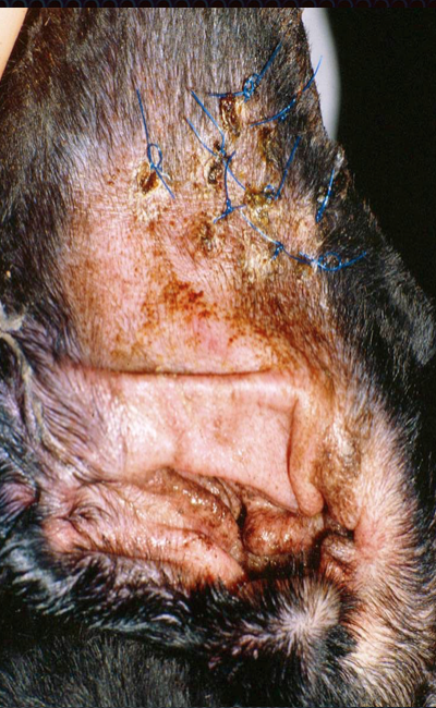
Postoperatively, place a stockinette on the dog’s head to protect the pinna and reduce bleeding. I prefer not to send dogs home with a full bandage on the ear or head. Keep the dog from scratching the ear with an Elizabethan collar. Remove sutures at 14 days.(Fig. 6)
Fig. 6: Pinna of a dog 2 weeks after the punch technique
for aural hematoma (Photo courtesy of Dr. Daniel Smeak)
If otitis externa or other skin disorder is present, treat appropriately.
References

1. Pavletic MM Use of laterally placed vacuum drains for management of aural hematomas in five dogs. J Am Vet Med Assoc. 2015 Jan 1;246(1):112-7.
2. Dye TL, Teague HD, Ostwald DA Jr, Ferreira SD. Evaluation of a technique using the carbon dioxide laser for the treatment of aural hematomas. J Am Anim Hosp Assoc. 2002 Jul-Aug;38(4):385-90.
3. Kagan KG Treatment of canine aural hematoma with an indwelling drain. J Am Vet Med Assoc. 1983 Nov 1;183(9):972-
4. Smeak DD. Surgery of the ear canal and pinna. Saunders Manual of Small Animal Practice, 3rd ed., Birchard and Sherding editors, Elsevier, 2006, pg. 582)

Blog Update: Dr. Birchard has published a new book: "Their Tails Kept Wagging", a collection of moving stories about pets with serious illness who survived. Click here for more information. 


Wednesday, June 10, 2015

Total Ear Canal Ablation in Dogs and Cats: A big surgery with big benefits.

Total ear canal ablation is removal of the entire ear canal. Indications for TECA include severe chronic otitis with obstruction of the canal, ear canal or middle ear neoplasia, severe trauma to the ear canal, or congenital abnormality that causes obstruction of the canal.

Key Point: Chronic proliferative otitis can result in calcification of the ear canal. This is an irreversible, end stage change in the ear that can only be resolved by TECA.

TECA is combined with a lateral bulla osteotomy (BO) to remove residual epithelium and debris from the middle ear after the ear canal is removed. Because of the prevalence of severe ear canal disease in dogs and cats, TECA/BO has become a common surgical procedure. However, a properly performed TECA/BO is a difficult procedure and can be associated with many complications. It should be performed by a board certified surgeon who is familiar with the anatomy of the ear and the technical aspects of the procedure. However, a well performed TECA can significantly improve quality of life of animals with ear disease.

Anatomy
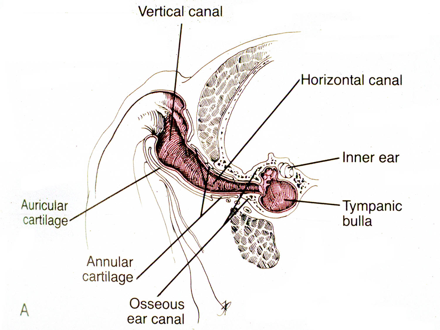
The entrance of ear canal, the external acoustic meatus, is surrounded by several cartilaginous structures including the tragus, antitragus, helix, and antihelix. The external ear canal in dogs and cats is divided into vertical and horizontal portions. (Fig. 1) 
Fig 1: Cross section of the ear canal in a dog.
(from: Smeak DD. Surgery of the ear canal and pinna. Saunders Manual of Small Animal Practice, 3rd ed., 
Birchard and Sherding editors, Elsevier, 2006)
The auricular cartilage is the vertical portion of the canal. The annular cartilage is located where the vertical canal turns into the horizontal canal. (Fig. 1)

The epidermal lining of the ear canal is rich in sebaceous and apocrine glands. In dogs with chronic otitis externa, an epithelial pouch may develop just adjacent and ventral to the tympanic bulla. (Fig. 2) 
Fig 2: Cross section of the ear in a dog with chronic otitis. Note the epithelial pouch
that develops in the canal adjacent to the entrance to the tympanic bulla. The tissue
colored red indicates that removed during a TECA/BO
(from: Smeak DD. Surgery of the ear canal and pinna. Saunders Manual of Small Animal Practice, 3rd ed.,
Birchard and Sherding editors, Elsevier, 2006)
The V-shaped parotid salivary gland lies over the ventro-lateral aspect of the vertical ear canal.

The middle ear is located in the petrous temporal bone. (Fig. 2) The tympanic bulla is the ventral wall of the tympanic cavity. It is an air-filled cavity just medial to the tympanic membrane. In the cat a septum divides the bulla into dorsomedial and ventrolateral compartments. Sympathetic nerve fibers run through the middle ear, and adjacent to the tympanic bulla are the facial nerve ventrolaterally, the carotid artery medially, and the hypoglossal nerve ventrally.

The major blood supply to the ear is via the great auricular artery and vein. Another important regional structure is the facial nerve. The nerve exits the skull just caudal to the ear canal and courses ventrally below the canal, then cranially. (Fig. 3) 
Fig. 3: Anatomy of important structures associated with the ear canal.
(from: Smeak DD. Surgery of the ear canal and pinna. Saunders Manual of Small Animal Practice, 3rd ed.,
Birchard and Sherding editors, Elsevier, 2006)
The nerve is motor to the lips and eyelids, therefore trauma to it causes lip droop and inability to blink.

Preoperative Considerations
Besides routine preoperative diagnostics such as history, physical examination, and blood tests, a good otoscopic exam and diagnostic imaging should be obtained on animals being considered for ear canal ablation.  Foreign bodies, neoplasia, or obstructive disorders of the canal may be discovered on otoscopic exam. Animals with tumors should also be screened for metastatic disease with thoracic radiographs, fine needle aspirate of regional lymph nodes if enlarged, and other tests as indicated. Skull radiographs or CT scan is usually recommended before TECA/BO to assess the tympanic bulla. See Veterinary Key Points blog “Nasopharyngeal Polyps in Cats”, 4/15/2015 for more discussion of bulla imaging techniques.

Surgical Procedure                      
After making the initial incisions around the external acoustic meatus and then ventrally along the vertical canal, carefully dissect the ear canal from surrounding tissues. (Fig. 4)
Fig. 4: Initial dissection in a canine cadaver for a TECA/BO. (note: Figures 4,5,7, and 8 are cadaver specimens.
 The cartilage around the external acoustic meatus has been severed 
and lifted up to allow dissection down the canal.
Dissect soft tissues close to the canal to avoid trauma to important structures, such as the facial nerve. (Fig. 5) 
Fig. 5: Continued dissection of the canal exposing the facial nerve in the stay suture. (arrow)
In ossified canals the facial nerve may be imbedded in the outer layer of the ear canal. (Fig. 6)
Fig. 6: The facial nerve (surrounded by yellow vessel loops) is being released from its adherence
to the ossified ear canal (arrow). Note the groove left in the canal by the nerve after gently dissecting it off.
 After removal of the canal using scalpel or scissors, carefully remove any remnants of canal and epithelium from the typanic bulla. Perform a bulla osteotomy with rongeurs to better expose the interior of the bulla. (Fig. 7) 
Fig. 7: After removal of the canal, the tympanic bulla is exposed by removing some of the
lateral aspect of the bony wall with rongeurs. Any remnants of the ear canal attached to the bulla
can also be removed with the rongeurs
Use a bone curette to remove epithelium and debris from the interior of the tympanic bulla. (Fig. 8) 
Fig. 8: Curettage of the interior of the bulla with a bone curette.
Avoid curettage of the dorsal aspect of the bulla to prevent trauma to the structures of the inner ear. Submit samples of fluid or debris from the tympanic bulla for culture and sensitivity. Also, submit the ear canal for histopathology to rule out neoplasia. Flush the incision with warm, sterile saline prior to closure. Close the incision in a “T” shape in multiple layers: deep fascia, subcutaneous tissue, and skin.

Postoperative Care and Complications
Postoperatively, protect the incision with a light bandage or Elizabethan collar. Administer analgesics for at least 3-5 days postoperatively. Long-term antibiotics (i.e. 3-4 weeks) are indicated in animals with bacterial infections. Choose antibiotics based upon the results of culture and sensitivity. If the animal’s eyelid motor function is decreased due to facial nerve injury, keep the eye lubricated with eye ointments or drops (e.g. Duratears) administered every 4-6 hours to prevent corneal ulcers until facial nerve function returns. 

Complications of TECA include acute pharyngeal edema, facial nerve damage, wound infection or dehiscence, Horner's syndrome, or deep abscesses. Deep abscesses occur due to leaving small amounts of secretory epithelium in or around the tympanic bulla. Reoperation to retrieve the residual epithelial tissue is usually necessary. Depending on the study, facial nerve deficits after TECA/BO in dogs can range from 36 to 48%, and in cats as high as 56%.(1-3) Although hearing is certainly diminished, some studies have found that some ability to hear is preserved even after removal of the ear canal.(2)

References
1. DD Smeak, WD DeHoff. Total Ear Canal Ablation Clinical Results in the Dog and Cat. Veterinary Surgery Volume 15, Issue 2, pages 161–170, March 1986
2. R. A. S. White, C. J. Pomeroy. Total ear canal ablation and lateral bulla osteotomy in the dog  Journal of Small Animal Practice Volume 31, Issue 11, pages 547–553, November 1990
3. Rebecca E. Spivack, A. Derrell Elkins, George E. Moore, and Gary C. Lantz (2013) Postoperative Complications Following TECA-LBO in the Dog and Cat. Journal of the American Animal Hospital Association: May/June 2013, Vol. 49, No. 3, pp. 160-168

Sunday, April 12, 2015

Nasopharyngeal Polyps in Cats: Key words - stertor, traction, and bulla osteotomy

Nasopharyngeal (NP) polyps in cats are characterized by well-circumscribed solid masses that are found in the nasopharynx, tympanic bulla, and Eustachian tube.  They are benign and thought to be secondary to inflammation from bacterial or viral infections.  Similar polyps can also be present in the external ear canal.  Affected cats are usually young (less than 1 year) but all ages can be affected.

Clinical Signs

Presenting signs of nasopharyngeal polyps may vary depending on location. Inspiratory stertor is commonly found due to the fleshy mass just dorsal to the soft palate causing obstruction of the nasal passages. Stertorous breathing in cats should alert the clinician to a mass lesion in the nasopharynx since other causes, such as elongated soft palate, are uncommon in cats.

Signs of otitis media, such as Horner’s syndrome, head tilt, and pain may also be seen. (Fig. 1) 
Fig. 1: Right sided Horner's syndrome seen in a kitten with a
NP polyp. 
Polyps in the external ear canal can result in or be secondary to otitis externa causing head shaking, ear scratching, and malodorous otic discharge.

Diagnosis

A thorough oral examination should be performed. Sedation will probably be necessary since the polyp will likely be obscured by the soft palate. If the mass is large enough simple palpation of the soft palate with a finger may indicate a mass effect. Retraction of the soft palate with stay sutures or a spay hook, with the cat in dorsal recumbency, should expose the mass. (Fig. 2)
Fig. 2: NP polyp in a cat (arrow) adjacent to the soft palate (SP) The cat is in
dorsal recumbency.

Otoscopic examination should also be performed to look for extensions of polyps into the external ear canal. Cleaning of otic discharge and debris may be necessary to adequately expose the mass.

Differential diagnoses for nasopharyngeal polyps are lymphosarcoma, other types of  neoplasia, foreign body, or congenital anomaly of the pharynx or upper airway. In one study of nasopharyngeal disorders in 53 cats 49% of them were diagnosed with lymphosarcoma and  28% were diagnosed as polyps.(1)

Imaging

Skull radiographs with emphasis on the tympanic bulla may provide useful information. Affected cats may show radiographic signs of chronic otitis media such as bony proliferation of the bulla and increased soft tissue density within the bulla.(Fig. 3)
Fig. 3: Bony proliferation of the bulla (arrow) secondary to
a nasopharyngeal polyp in a cat.
However, radiographs of the bullae are not a very sensitive imaging test and significant changes may be present with normal appearing bullae on films.

CT scans are a more sensitive and diagnostic imaging modality for bulla disorders and are useful in cats with nasopharyngeal polyps or other bulla diseases. Increased soft tissue density is commonly seen in one or both bullae. (Fig. 4)
Fig. 4: CT scan of a cat with a NP polyp showing increased soft tissue density
within the bulla. (arrow)
In cases of severe infection or invasive neoplasia, lysis of the bulla may be seen. These findings are useful for preoperative evaluation of cats undergoing ventral bulla osteotomy, a recommended surgery for cats with nasopharyngeal polyps or other chronic middle or external ear disease.

Treatment

Traction

Surgical removal of the polyps is the most effective means of relieving clinical signs. Under general anesthesia the cat is placed in dorsal recumbency and a mouth speculum placed to allow exposure of the oral cavity and pharynx. The soft palate is retracted rostrally with either stay sutures or a spay hook. Gentle manipulation of this tissue is important since it is sensitive and prone to edema with manipulation.  Even under anesthesia many cats will exhibit discomfort while the soft palate is being manipulated. Once the polyp is exposed, grasp it with either stay sutures or Allis tissue forceps.(Fig. 5) Gentle but firm rostral and ventral traction is placed on the polyp and continued until the mass is removed. 
Fig. 5: NP polyp (arrow) being removed by traction.
The tissue frequently has a large round component that is connected to a tail that is the Eustachian tube portion of the polyp. Control hemorrhage with direct pressure on the affected pharyngeal tissues.

Bulla Osteotomy

To completely remove the polyp tissues, ventral bulla osteotomy is then performed. Which side to operate depends on the preoperative imaging or lateralizing clinical signs. If lateralization was not possible or if changes are seen bilaterally on imaging, both bullae are ostetomized to be sure of removing all remaining polyp tissues. Although most polyps are unilateral, the author recently treated a cat with bilateral polyps requiring osteotomy of both bullae.

Small fragments of polyp tissue are usually found in the tympanic bullae exposed by the ventral bulla osteotomy. (Fig. 6-7)
Fig. 6: Diagram of a ventral bulla osteotomy in a cat. Rongeurs are used to
remove the ventral aspect of the bulla and expose both compartments.
from: Boothe H. Surgery for otitis media and otitis internal.
From: Saunders Manual of Small Animal Practice3rd ed., Birchard and Sherding, editors. Elsevier, 2006, pg. 601.

Fig. 7: Bulla osteotomy in a cat with NP polyps. The ventral floor of the
bulla has been removed. Note the polypoid tissue filling the bulla cavity. (arrow)

It is important to expose both compartments of the bulla when performing the osteotomy (ventral-medial and dorso-lateral chambers). Care is taken not to injure components of the inner ear, located dorsally in the bulla, when doing polyp excision and curretage.  Save all tissues removed for histopathology and obtain samples from the bulla for culture and sensitivity.

Postoperative Care and Complications

Routine supportive care including analgesics and antibiotics are administered postoperatively. Antibiotic choice is guided by results of culture and sensitivity of samples obtained from the bullae.

Common complications after polyp removal are pharyngeal swelling and Horner’s syndrome due to injury to sympathetic nerves in the middle ear. Both of these problems are usually mild, short term in duration, and not requiring treatment. Recurrence of polyps is more likely if only the nasopharyngeal portion is removed. In a clinical study of 31 cats with nasopharyngeal polyps, 5 recurred postoperatively, 4 of which did not have a bulla osteotomy. (2)

References

1. HS Allen, J Broussard, and K Noone (1999) Nasopharyngeal diseases in cats: a retrospective study of 53 cases (1991-1998). Journal of the American Animal Hospital Association: November/December 1999, Vol. 35, No. 6, pp. 457-461. 
2. Kapatkin, AS, Matthiesen, DT, Noone KE. et.al. Results of surgery and long-term follow-up in 31 cats with nasopharyngeal polyps. J Am An Hosp Assoc 1990 Vol 26 No 4 pp. 387-392.