Author

Stephen J. Birchard DVM, MS, Diplomate ACVS

Saturday, March 28, 2026

Demystifying Bandage Selection for Wounds in Dogs and Cats

In treating open skin wounds in small animals, one of the most important decisions you make is bandage selection. It’s easy to think of bandaging as a routine step after wound preparation, but in reality, the bandage plays an active role in how the wound heals. The right bandage can help debride, control infection, manage exudate, and protect developing tissue. The wrong bandage — or the right bandage used at the wrong time — can delay healing and create new problems.

Over the years, I’ve found that many clinicians don’t struggle with the concept of wound management as much as they struggle with choosing the most appropriate bandage for the situation at hand. There are many options available, and it’s not always obvious which one to use — especially in the early stages of wound care.

The goal of this post (Part 1) is to provide a simple, practical reference to commonly used bandage types for open wounds. This is not meant to be exhaustive, but rather a guide you can quickly review and apply in everyday practice. Stay tuned for Part 2 which will address in which wound phase to use these bandage types. 

Table: Bandage Types and Indications

Wet-to-Dry Bandage – Adherent, provides mechanical debridement of necrotic tissue; used in contaminated or necrotic wounds early in management. 

Tie-Over Bandage – Provides a stable dressing in difficult locations such as the axilla, groin, and head; useful for a variety of bandage primary layers. (See here for more information)

Non-Adherent Contact Layer (e.g., Adaptic Touch) – Protects granulation tissue; used for fresh wounds not needing debridement, graft beds, and partial-thickness injuries. (See here for more information)

Sugar or Honey Dressings – Bactericidal, absorbent, non-adherent; used in infected wounds to reduce bacterial load and support early granulation. (See here for more information)


Petroleum-Impregnated Gauze – Used on well developed granulation tissue that is clean and not infected, non-adherent. (See here for more information)


Telfa Pad – Non-adherent, minimally absorbent; used for free skin grafts or closed incisions (not used on open wounds).

Topical Antibiotic Ointment – Protect closed incisions and small superficial wounds; not a substitute for systemic therapy in deep infections.

Successful wound management depends not only on choosing the right bandage, but on understanding what that bandage is meant to accomplish. Each layer has a purpose, and using them thoughtfully can make the difference between delayed healing and an efficient, uncomplicated recovery.

In Part 2, we’ll take the next step—matching bandage selection to the phase of wound healing, so you can make practical, case-based decisions in real time.

The concepts and tables presented here are adapted from our upcoming textbook on small animal soft tissue surgery, currently in development as a practical, quick-reference resource for clinicians.


Monday, January 26, 2026

Inspiratory Stridor in an Older Labrador: Is It Laryngeal Paralysis?

A 10-year-old neutered male Labrador retriever was referred for evaluation of progressive inspiratory dyspnea of several weeks’ duration, with marked worsening over the preceding 24 hours. The dog also had a one-week history of anorexia and had lost approximately 3 kg. Oral examination by the referring veterinarian raised concern for a possible laryngeal mass, prompting referral. 

On presentation, the dog was alert and ambulatory but exhibited moderate inspiratory stridor. He was mildly dehydrated and thin, with a body condition score of 2/5. Results of a complete blood count, serum chemistry profile, and thyroid testing (TSH and free T4 by equilibrium dialysis) were within normal limits. Thoracic radiographs were unremarkable aside from mild degenerative joint disease of the shoulders. Given the dog’s signalment and clinical signs, laryngeal paralysis was considered the leading differential diagnosis. 

To further evaluate laryngeal function, IV propofol was administered to permit a thorough laryngeal examination. Arytenoid function appeared normal, with adequate abduction bilaterally. However, distortion of the mucosa ventral to the epiglottis was observed, along with a pedunculated bridge of mucosa connecting to a mass located to the left of the larynx and extending into the proximal esophagus. 

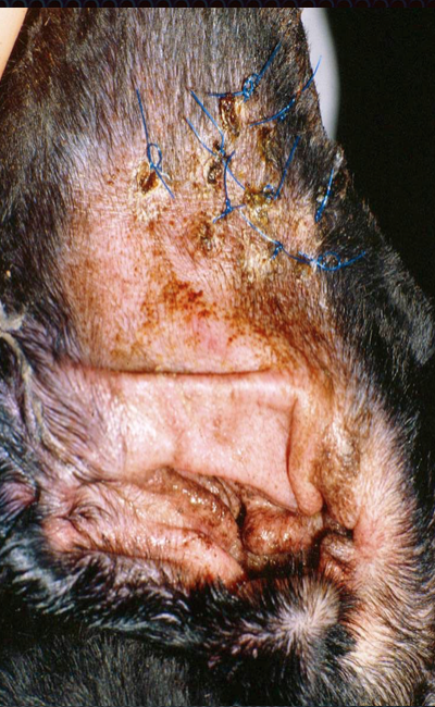
General anesthesia was then induced and maintained with isoflurane via a cuffed endotracheal tube. The dog was positioned in sternal recumbency, and Babcock forceps were used to gently retract the mass into the pharynx. The mass was cylindrical, approximately 7 cm long and 4 cm wide (Fig. 1, 2). 

Fig. 1: Oral mass in oral cavity (held by forceps)


Fig. 2: Pedunculated portion of mass (white arrow)

The mucosal peduncle was ligated using one circumferential and one transfixing ligature of 2-0 polydioxanone, followed by sharp excision with Metzenbaum scissors. A small residual mucosal defect ventral to the epiglottis was closed with a single simple interrupted suture. No additional abnormalities or hemorrhage were noted on repeat laryngeal examination. Fine-needle aspirates of the submandibular lymph nodes, which were normal in size, were obtained prior to recovery. Recovery from anesthesia was uneventful. 

The dog was discharged two days postoperatively, breathing comfortably and able to eat and drink without difficulty. Cytologic evaluation of the lymph nodes was unremarkable. Histopathology of the excised mass revealed a high-grade neurofibrosarcoma, with complete surgical excision. At recheck 10 days postoperatively, the owner reported marked improvement. The dog had no difficulty breathing, swallowing, or eating, and inspiratory stridor had resolved completely. Three months after surgery, the dog continued to do well, had gained weight, and remained clinically normal. Although consultation with an oncologist was recommended, the owner declined adjunctive chemotherapy or radiation therapy. 

Discussion 

This case represents an unusual cause of inspiratory stridor and weight loss in an older Labrador retriever. The initial presentation was highly suggestive of laryngeal paralysis, emphasizing the importance of maintaining a broad differential diagnosis in dogs with upper airway obstruction. Sedated laryngeal examination was essential in establishing the correct diagnosis. In addition to respiratory signs, this dog had a notable history of anorexia and weight loss. The pedunculated nature of the mass allowed it to intermittently prolapse into the proximal esophagus, likely interfering with normal swallowing despite the absence of overt dysphagia reported by the owner. Advanced imaging of the pharyngeal and laryngeal region may have aided in identifying a mass; however, the mobility of the lesion makes detection by plain radiography unlikely unless contrast material is used. Computed tomography or ultrasonography may be useful in selected cases, and ultrasonography has been described for evaluation of laryngeal disorders in dogs [1]. 

Neurofibrosarcomas, also classified as peripheral nerve sheath tumors, are rare in the oral cavity or pharynx of dogs. These tumors more commonly arise from peripheral nerves, the brain, or spinal cord and are considered a subtype of soft tissue sarcoma. Reported oral tumors in dogs more frequently include melanoma, squamous cell carcinoma, and fibrosarcoma. Laryngeal tumors themselves are uncommon but encompass a wide range of histologic types [2,3]. The tumor in this case was unusual not only for its histologic classification but also for its location and pedunculated morphology, which allowed complete excision without removal of any laryngeal cartilages. In contrast, most laryngeal tumors are not amenable to complete resection without aggressive procedures such as partial or total laryngectomy. High-grade soft tissue sarcomas carry a guarded long-term prognosis due to the risk of local recurrence and metastasis [4]. More recent studies suggest improved outcomes when surgery is combined with radiation therapy [5]. Early detection and complete surgical excision remain critical, and consultation with an oncologist should be considered when feasible. 

Summary 

Not all Labradors with inspiratory stridor have laryngeal paralysis. Careful diagnostic evaluation, including sedated laryngeal examination, is essential before committing to definitive airway surgery. This case highlights the value of surgical judgment, restraint, and thorough assessment in achieving an excellent outcome. 

 References
 
1. Rudorf H, Barr FJ, Lane JG. The role of ultrasound in the assessment of laryngeal paralysis in the dog. Vet Radiol Ultrasound. 2001;42(4):338–343. 
2. Ruppert C, Hartmann K, Fischer A, et al. Cervical neoplasia originating from the vagus nerve in a dog. J Small Anim Pract. 2000;41(3):119–122. 
3. Withrow SJ, Vail DM, Page RL. Small Animal Clinical Oncology. 
4. Ciekot PA, Powers BE, Withrow SJ, Straw RC, Ogilvie GK, LaRue SM. Histologically low-grade, yet biologically high-grade, fibrosarcomas of the mandible and maxilla in dogs: 25 cases (1982-1991). J Am Vet Med Assoc. 1994 Feb 15;204(4):610 
5. McKnight JA, Mauldin GN, McEntee MC, et al. Radiation treatment for incompletely resected soft-tissue sarcomas in dogs.

Monday, May 19, 2025

The Punch Technique for Aural (Ear) Hematomas in Dogs and Cats: Simple, effective, and cosmetic!


Auricular hematomas occur from hemorrhage that develops between the 2 leaves of cartilage of the pinna. The hemorrhage can occur due to trauma to the pinna from head shaking or scratching.  Inflammatory conditions of the ear canal, such as ear mites, foreign bodies, or bacterial otitis may be the inciting cause.

Diagnosis

Aural hematomas are characteristic in appearance.(Fig.1,2) 

Fig. 1: Aural hematoma in a cat
Fig. 2: Aural hematoma in a young white tiger

The pinna is enlarged and fluctuant. Differential diagnoses include acute allergic response causing severe swelling of the pinna, and neoplasia of the pinna. Aspiration of the mass reveals blood or serum. Rule out underlying ear canal problems by a thorough palpation of the ear canal and otoscopic exam. Also, thoroughly examine the animal for evidence of skin disease such as allergies, seborrhea, fleas, or pyoderma. 


Treatment

Many methods have been described for treatment of aural hematomas. Incision and drainage, drain tubes, and laser techniques have all been described.(1-3) Medical management by simple drainage combined with either systemic or local corticosteroid therapy has also been advocated. The advantage of medical therapy or simple needle drainage is excellent cosmetic result. However, incidence of recurrence with these treatments is high. The advantage of incision and suture is a low rate of recurrence, but the scaring of the pinna can cause poor cosmetic results.

The punch technique described here (Fig. 3) allows effective drainage and very low incidence of recurrence.(4) The cosmetic results are also very good since little scar tissue develops in the small incisions.
Fig. 3: Depiction of punch technique for aural hematomas in dogs and cats.
(from: Smeak DD. Surgery of the ear canal and pinna. Saunders Manual of Small Animal Practice, 3rd ed.,
Birchard and Sherding editors, Figure 60-1, Elsevier, 2006, pg. 583)

Surgical Technique
  • Clip and prepare both sides of the pinna for aseptic surgery. Place a surgical sponge in the ear canal to prevent accumulation of blood.
  • Use a skin biopsy punch (size 4-6 depending on the size of the dog) to remove small plugs of skin and cartilage on the medial side of the pinna.(Fig. 4) 
    Creating punch incisions on the medial aspect of the pinna for  drainage of aural hematoma.
  • Attempt to penetrate only the skin and 1 layer of the cartilage with the punch; however inadvertent removal a small section of both of the cartilage layers is not problematic.
  • Make incisions about 0.5 – 1 cm apart and perform as many punches as necessary to drain the entire hematoma. 
  • Tack the skin edge of each incision with monofilament nylon, polypropylene, or Monocryl in a simple interrupted pattern.(Fig. 5) The size of suture can be 3-0 or 4-0 depending on the size of the animal. It is not necessary for the suture to penetrate full thickness through all layers of the pinna including the skin on both sides but the suture should incorporate both layers of cartilage and the skin on the medial surface.

Fig. 5: Suturing the edge of each punch incision with monofilament suture.

Postoperative Care

Postoperatively, place a stockinette on the dog’s head to protect the pinna and reduce bleeding. I prefer not to send dogs home with a full bandage on the ear or head. Keep the dog from scratching the ear with an Elizabethan collar. Remove sutures at 14 days.(Fig. 6)
Fig. 6: Pinna of a dog 2 weeks after the punch technique
for aural hematoma (Photo courtesy of Dr. Daniel Smeak)
If otitis externa or other skin disorder is present, treat appropriately.

References

1. Pavletic MM Use of laterally placed vacuum drains for management of aural hematomas in five dogs. J Am Vet Med Assoc. 2015 Jan 1;246(1):112-7.
2. Dye TL, Teague HD, Ostwald DA Jr, Ferreira SD. Evaluation of a technique using the carbon dioxide laser for the treatment of aural hematomas. J Am Anim Hosp Assoc. 2002 Jul-Aug;38(4):385-90.
3. Kagan KG Treatment of canine aural hematoma with an indwelling drain. J Am Vet Med Assoc. 1983 Nov 1;183(9):972-
4. Smeak DD. Surgery of the ear canal and pinna. Saunders Manual of Small Animal Practice, 3rd ed., Birchard and Sherding editors, Elsevier, 2006, pg. 582)

Blog Update: Dr. Birchard has published a new book: "Their Tails Kept Wagging", a collection of moving stories about pets with serious illness who survived. Click here for more information. 


Sunday, May 18, 2025

Scrotal Hematoma in Dogs After Castration: Why does it happen and how to prevent it.

Scrotal hematoma, the accumulation of blood in the scrotal sac, is a common postoperative complication of castration in dogs. (Fig. 1) 
Fig. 1: Scrotal hematoma that developed after castration in this golden retriever
Postoperative bleeding from one or both testicular arteries or veins is the most likely cause. Bleeding from these vessels can also accumulate in the abdominal cavity and be unrecognized until the dog becomes hypovolemic and develops shock. Bleeding from subcutaneous tissues could also result in a scrotal hematoma but is less likely, particularly in cases where bleeding is severe and the scrotal hematoma is large.

Etiology
Insecure ligatures are probably the cause of bleeding from testicular vessels. Ligature failure during castration may be due to a number of technical problems, such as loose ligature, improper suture material, or ligature slippage. Improper location of the suture ligature can also predispose to failure. If the spermatic cord is not adequately exteriorized the ligatures will be placed too close to the testicle. (Fig. 2) 
Fig. 2: The red dotted line indicates ligation of the spermatic cord too close to the testicle (T)
The ligatures in the figure are placed in the proper location.
(Modified from: Boothe, HW. Surgery of the testicles and scrotum. Saunders Manual of Small Animal Practice, Birchard and Sherding editors,  Figure 87-1, Elesevier, 2006, pg. 970.)
This causes excessive tissue to be incorporated in the ligatures. Insufficient stripping of the adipose tissue from the cord also predisposes the ligature to slippage.

Treatment
Scrotal hematomas can be managed medically with compresses (ice packs for the first 24 hours postoperatively, followed by warm compresses), Elizabethan collar, restricted exercise, analgesics such as NSAIDS, and antibiotics. In severe cases or those not responding to medical therapy, scrotal ablation is necessary to remove the entire scrotum and its contents.

Prevention
To prevent scrotal hematoma when performing castration, adhere to the following technical principles:
- After incising over the testicle and exposing it, break down the gubernaculum testis and place traction on the testicle to allow several centimeters of the spermatic cord to be exposed. (Fig. 3)
Fig. 3: Proper exposure and clamping of the spermatic cord during routine canine castration.
- Carefully and thoroughly strip the adipose tissue from the spermatic cord with a moistened surgical sponge.
- Triple clamp the cord with Carmalt clamps, cut between the 2 clamps closest to the testicle and perform routine ligatures with Monocryl or PDS suture (transfixing and full ligatures). The full ligature is placed in the crush area of the most proximal clamp after it is removed, the transfixing ligature is placed just distal to the full ligature. (Fig. 4)
Fig. 4: Proper ligature technique in a closed canine castration. (T=testicle)
(Modified from: Boothe, HW. Surgery of the testicles and scrotum. Saunders Manual of Small Animal Practice, Birchard and Sherding editors, Elesevier, 2006, Figure 87-1, pg. 970.)
- Check the vascular stump for bleeding prior to replacing it into the incision. Repeat the same procedure on the opposite side.  

I prefer closed castration, even on large dogs, because it allows excision of the testicular tunics along with the testicle. (Fig. 5)
Fig. 5: Closed castration allows removal of the parietal vaginal tunic with the testicle
Close the subcutaneous tissue routinely (3-0 or 4-0 absorbable suture in a simple continuous pattern). Although skin sutures can be used on the skin incision, I prefer do an intradermal closure in lieu of skin sutures. Fewer complications occur with an intradermal closure of castration incisions. In an unpublished study performed at Ohio State several years ago, dogs with intradermal skin closure had fewer incisional problems and even fewer scrotal hematomas postoperatively. Less self-trauma to the incision and scrotum were thought to be the reason for this finding.

__________________________________________________________________


When seriously ill pets are surrounded with love, they are more likely to survive. Read about this in Dr. Birchard's new book: "Their Tails Kept Wagging". Click here for more information.

Friday, February 9, 2024

Introducing the Air Plasma Surgical Device (Guest Blogger Dr. William T. N. Culp)


William T. N. Culp, DVM, Diplomate, ACVS

Surgery & Oncology Department 
Veterinary Teaching Hospital
UC Davis School of Veterinary Medicine
Davis, California


AirPlasma is a recently developed technique in which air is ionized to generate energy. This process is conducted without the use of gases such as argon and helium and utilizes only air as a conductor. The energy that is created is visible in the form of a glow and tissue can be vaporized with simultaneous capillary hemostasis. (Lacitignola 2020)

AirPlasma has several advantages over other forms of similar devices. The AirPlasma device works at a much lower temperature than other plasma devices, and the local thermal damage at the point of contact can be minimized with significantly less tissue necrosis. AirPlasma does not require a return plate on the patient which avoids burns on the patient’s skin encountered with some electrosurgical devices. There also is evidence that AirPlasma has a low carcinogenic potential which is particularly important as other devices can generate fumes that result in exposure to certain carcinogens. Lastly, as stated above, AirPlasma devices are able to generate energy without the need for external gases.

There are currently 3 major areas where AirPlasma is utilized in veterinary patients: ablation of tumors, cutting, dissection, and micro-coagulation. Small masses can be ablated with the device utilizing several available tip options. The device is particularly useful for cutting and dissecting tissues as the generated plasma can be highly focused on the targeted area. Because of this advantage, cutting and dissection during procedures such as total ear canal ablation, staphylectomy, and tumor removal can be very precise with minimal trauma to surrounding tissues. Finally, this device is particularly useful in assisting with microcoagulation at surgical sites after biopsies or resections. Commonly, after liver lobectomy or other partial organ resection, a small amount of bleeding can occur. Using the AirPlasma on those sites can often control minor oozing and prevent continued blood loss.

AirPlasma technology is an exciting new modality that is demonstrating promise. Proficiency is achievable in a short period of time, and safety appears to be high. Further studies will continue to elucidate outcomes with this technology, and the applications in veterinary patients are likely to increase. For more information about the AirPlasma device, and Onemytis see http://www.medviatech.com/.


Reference

Lacitignola L, Desantis S, Izzo G, et al. Comparative morphological effects of cold-blade, electrosurgical, and plasma scalpels on dog skin. Vet Sci 2020;7,8; doi:10.3390/vetsci7010008 


Monday, September 4, 2023

Torsion of a Cryptorchid Testicle


Tank is a 5-month-old male intact boxer who presented to the Emergency Service for acute vomiting and abdominal pain. The vomiting began about 12 hours before presentation. The referring veterinarian had obtained abdominal radiographs and was suspicious of a gastric foreign body. There was no other history of medical problems with Tank.


On physical examination, Tank was quiet but responsive, ambulatory, and mildly dehydrated. He was painful on abdominal palpation, especially in the mid to caudal abdomen. Only one testicle was palpable in his scrotum, which was determined to be the right testicle. The left testicle was not palpable in the inguinal area.


A complete blood count and serum chemistry profile revealed no significant abnormalities. On review of the referring veterinarian's abdominal radiographs, we did not find evidence of a gastric foreign body or other abnormality. 


Abdominal ultrasound revealed a structure that appeared to be a testicle. (Fig 1)

Fig 1: Abdominal ultrasound showing a cryptorchid testicle (arrow)


Placement of the ultrasound probe on the structure elicited severe pain. We were concerned that Tank had torsion of the abdominal cryptorchid testicle. 


A left paramedian abdominal exploratory was performed. Torsion of the left retained testicle was confirmed, and the testicle was removed.

Fig. 2: Surgical removal of the abdominal testicle
 

The descended testicle was also removed. 

Fig. 3: Both testicles removed from Tank, normal on the right, testicular torsion on the left

Tank made an unremarkable recovery from anesthesia and surgery and was discharged from the hospital the following day. 


Cryptorchidism is a common congenital anomaly in male dogs. One or both testicles can be retained in the inguinal region, inguinal canal, or abdomen. For more details on surgery for this disorder, see https://drstephenbirchard.blogspot.com/2014/07/cryptorchidism-in-dogs-5-ways-to-make.html, and scan the QR code for a complete discussion of cryptorchidism in dogs and cats.


Sunday, February 19, 2023

Adaptic Touch: The Ideal Dressing For Open Skin Wounds in Dogs and Cats

When managing open skin wounds in dogs and cats, bandaging materials and techniques are critical elements of the treatment strategy. The interior of the bandage in contact with the wound is called the primary layer and is the most important of the dressing for creating a suitable environment for healing. Many options for the primary bandage layer are available, including wet saline sponges, dry sponges, silicone dressings, medical-grade honey gauze, petroleum-impregnated gauze, and Telfa pads. The ideal first layer of the bandage should encourage granulation tissue formation. It should not inhibit healing cells and not promote infection. The primary layer should allow absorption of wound exudate into the second layer of the bandage to prevent accumulation at the wound surface.  


Telfa pads have historically been used for open wounds in animals, but newer materials that allow better absorption of exudate from the wound are available. Wet saline sponges are commonly used as the initial primary bandage layer in acute traumatic wounds because they are highly absorptive, allow gentle debridement of dead tissue, and are inexpensive. However, when saline sponges become dry, they are adherent, making bandage changes painful. Patient sedation is frequently necessary to prevent discomfort during removal. Lavage of the dried sponges with sterile saline during bandage change will allow them to be peeled off the wound more easily and with less pain. Although wet saline sponges are very effective in initially managing severe wounds, such as bite wounds in dogs and cats, a primary bandage layer is needed for the next healing phase. Ideally, this material should allow absorption of wound exudate and be non-adherent to reduce patient discomfort. 


Vitalitymedical.com


The Adaptic Touch is an excellent choice for the primary bandage layer on open wounds in dogs and cats. It is a soft and pliable silicone-coated mesh material that readily conforms to the wound surface. The pore size in the mesh is small enough to prevent the ingrowth of granulation tissue into the material. The Adaptic Touch mesh allows the passage of wound exudate into the secondary bandage layer and is non-adherent, minimizing patient discomfort during bandage changes. It has been used extensively for various human wounds and is gaining favor with veterinarians. Cotton is a good choice for the secondary layer of the Adaptic Touch bandage allowing for the transmission of fluid through the pores of the mesh. Use only one layer of the Adaptic to ensure adequate absorption into the cotton.


The original Adaptic mesh is a petroleum-impregnated gauze that allows absorption and is non-adherent. However, petroleum may have some detrimental effects on wound healing, and it is only partially non-adherent. These characteristics make them less ideal than their counterpart, the Adaptic Touch.


See the above video for a demonstration of the non-adherent nature of the Adaptic Touch dressing. 


In summary, the ideal bandages for open skin wounds in dogs and cats have primary layers that allow absorption of wound exudate, are non-adherent, and do not inhibit wound healing. For wounds that are highly contaminated and filled with dirt and debris that cannot be entirely removed by surgical debridement and flushing, moist saline sponges are indicated as the initial bandage. Since these bandages are adherent and painful to remove, transition to non-adherent dressings such as the Adaptic Touch is recommended for the next phase of wound treatment.


References

International case series: Using ADAPTIC TOUCH® Non-Adhering Silicone Dressing: Case Studies. London: Wounds International, 2013. 

Wounds International case studies evaluation. ADAPTIC TOUCHTM Non-Adhering Silicone Dressing in skin tear management. Wounds International, 2018 (Suppl). Cooper 

Janice Bianchi*, Simon Barrett, Fania Pagnamenta, Fiona Russell, Sandra Stringfellow, Pam
Cooper. Consensus guidance for the use of Adaptic Touch non-adherent dressing. Wounds UK, 2011, Vol 7, No 3 
 

When surrounded with love, seriously ill pets are more likely to survive. Read about this in Dr. Birchard's new book. Click here for more information.