

Preoperative Care
Fluid
therapy is a critical aspect of overall management of the intestinal surgery
patient. Correct dehydration and provide maintenance needs with intravenous
fluids (such as Lactated Ringers or other balanced electrolyte solution). Correct
electrolyte abnormalities such as hypokalemia. Give colloids if the animal is
hypoproteinemic or whole blood or packed cells if anemic.
Prophylactic use of antibacterials is indicated since the
intestine is contaminated. Broad-spectrum antibiotics are recommended such as one of
the cephalosporins (cefazolin, 20 mg/kg IV) Begin the drug before surgery to
insure adequate blood levels at the time of the operation. Do not continue the
antibiotic postoperatively unless infection (e.g. peritonitis) is present.
Quietly recite the Halsted
Chant to prepare yourself for the surgery. (See first blog in the VKP series.)
Surgical Technique for Enterotomy
See previous blog on
Intestinal Biopsy for recommendations on instruments and sutures for intestinal
surgery. Use meticulous, atraumatic technique and keep the tissues moist.
Isolate the affected segment of bowel with moistened sponges and place stay
sutures adjacent to the proposed incision.
 |
| Fig. 1 Corn cob foreign body in the intestine of a dog. Make enterotomy incision at arrow. |
Have an assistant use their fingers to occlude the bowel on
each side of the enterotomy incision. Incise the intestine on the antimesenteric
side close to the foreign body and in an area of intestine that is healthy,
i.e. the area of bowel that is downstream from the foreign body. (Figs.1,2)
 |
| Fig 2: Fragment of rubber ball in the intestine of a dog. Make enterotomy incision at arrow. |
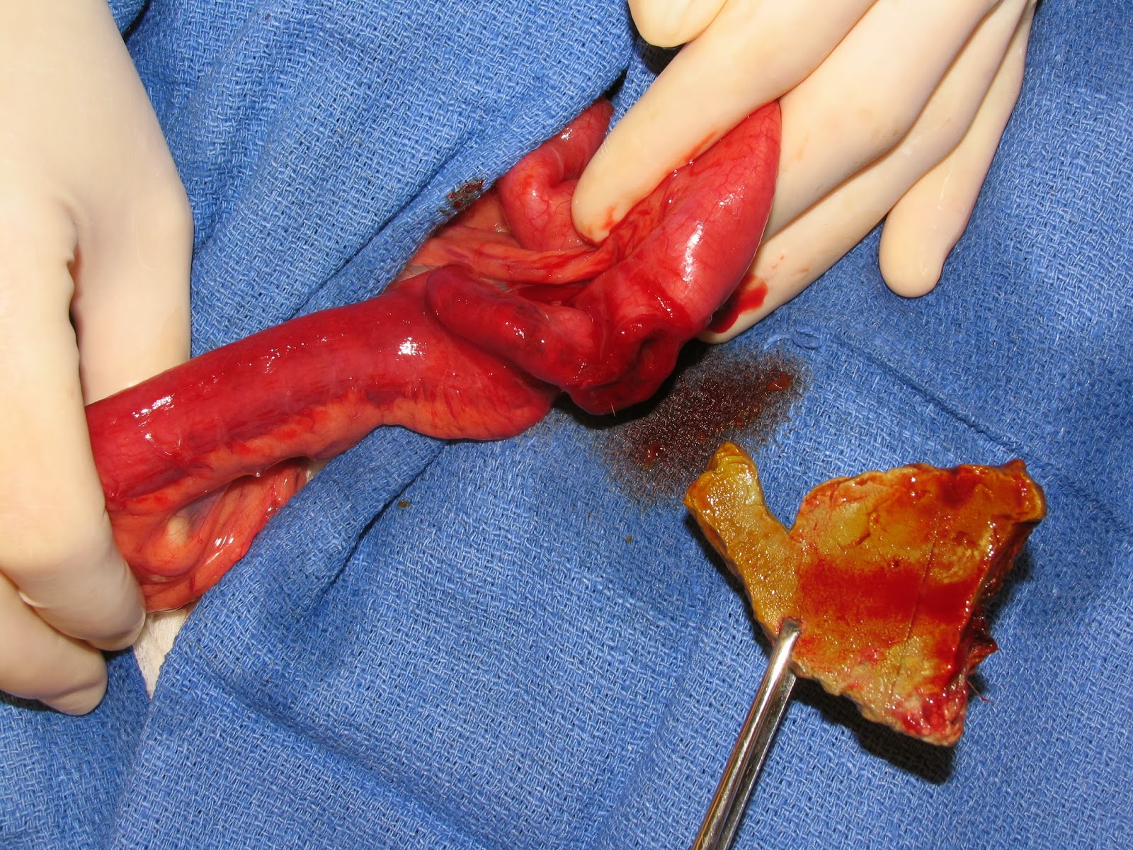 |
| Fig 3: Rubber fragment removed |
Use suction to control spillage and minimize contamination of the peritoneal cavity. Do not use electrocautery on the intestine for hemostasis. Extricate the foreign body from the intestine and remove it from the sterile field to avoid contamination. (Fig. 3)
Closure:
Close the intestine with 4-0
PDS, taper RB1 needle in a simple continuous pattern. Be sure to do full thickness bites of the intestine (Fig. 4) to include the submucosa which is the holding layer.
 |
| Fig. 4: Full thickness suture bites of the intestinal wall are required to be sure of including the submucosa. |
Avoid
excessively handling the full thickness bowel with thumb forceps even if you
are using DeBakey forceps. Have
your assistant maintain tension on the suture line as you are taking the bites
of tissues to keep it from loosening. Use the suture needle to guide each suture loop onto the incision to maintain even spacing. (See Video) After completing the simple continuous
line, fill in any gaps with the same suture using a simple interrupted pattern. (Fig. 5)
 |
| Fig. 5: Completed enterotomy closure with simple continuous pattern |
Leak test the incision as described in the blog on Intestinal Biopsy. Lavage
the local tissues but not the entire abdomen unless there was gross spillage of
intestinal contents or peritonitis is present. Place the omentum on the
incision and tack it adjacent to each end of the incision. (The omentum
helps seal the incision and provides blood supply and lymphatic drainage.)
See blog on Intestinal Biopsy
for postoperative care.
Always save the foreign body
and give it back to the owner when the animal is discharged from the hospital.
Encourage them to provide safe chew toys for their animal to use.
Video
The video is short but demonstrates some important principles of tissue handling that were mentioned in the text above.
No comments:
Post a Comment