
Elliptical incisions commonly
result from removal of skin tumors, other lesions, or debridement of traumatic wounds. Since loss of a section of
skin occurs, the closure of the defect can result in so called “dog-ears” at
each end of the incision (small flaps of skin that protrude from the ends of
the incision). An easy method to
prevent dog-ears and create a cosmetic and secure closure is the “rule of
halves” technique, also called bisectional closure. (Fig. 1)
 |
| Fig. 1: The rule of halves for closing elliptical incisions. Dotted lines represent placement of sutures |
This technique is used for
the deep fascia and subcutaneous layers of the wound. The sutures below the skin relieve
the tension across the incision and align the skin edges making the final
suturing of the skin much easier.
Technique for the Rule of Halves Closure
Take the first suture bite of the deep fascia and/or subcutaneous tissue in the middle of the incision. (Fig. 2)
 |
| Fig. 2: Surgical model of elliptical incision. The subcutaneous tissue is red, the skin is pink. Dotted line indicates where first subcutaneous suture is placed. |
This divides
the incision into 2 equal parts. (Fig. 3)
 |
| Fig. 3: First subcutaneous suture has been placed. Dotted lines indicate placement of the next 2 sutures. |
Now take suture bites in the middle
of each of the 2 defects, then in the middle of the 4 defects, and so on until
the subcutaneous layer is completely closed. (Fig. 4)
 |
| Fig. 4: Final subcutaneous suture being placed. Note that the suture knot is buried by taking the first bite, from inside out, on the side closest to the surgeon. |
Then suture the skin routinely. (Fig. 5)
 |
| Fig 5: Completed closure |
This technique for wound suturing,
although somewhat more time consuming than doing simple continuous patterns for
the subcutaneous layer, results in a very secure and cosmetic closure. (Figs. 6-8)
 |
| Fig. 6: Proposed lines for excision of a mast cell tumor in a dog. (X = tumor, circle indicates 3 cm margins, elliptical lines are proposed incision) |
 |
| Fig. 7: Same dog as in Fig. 6 after excision of the tumor and surrounding skin. Dotted line indicates location of first suture. |
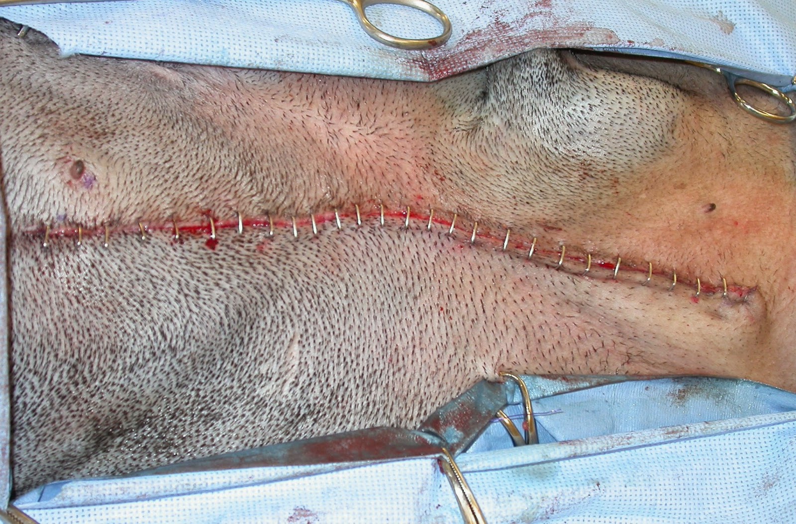 |
| Fig. 8: Completed closure of same dog in Fig. 6. |
No comments:
Post a Comment