 |
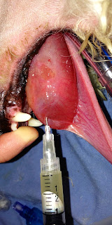
| Fig. 1: FNA of the oral mass in Ollie |
The gross appearance of the mass, its fluctuant nature, and the mucoid fluid that was aspirated (Fig. 1) were all suggestive of a rannula (oral mucocele). This is an accumulation of saliva from the sublingual salivary glands into the submucosal space in the mouth.
The treatment for Ollie was marsupialization of the rannula (Fig. 2-3), and removal of the mandibular and sublingual salivary glands. (Fig. 4)
 |
| Fig. 2: Completed marsupializaton of the rannula |
 |
| Fig. 3: Close up of the marsupialized rannula showing the interior of the mucocele. |
Marsupialization was performed by first incising over the dorsal aspect of the rannula with a scalpel. The saliva was evacuated from the cavity and the dorsal aspect of the rannula was debrided to make a large opening. The wall of the rannula was sutured to the inner tissue layer with 4-0 Monocryl in a simple continuous pattern.
The right mandibular and sublingual salivary glands were removed in the standard fashion through a lateral cervical incision directly over the mandibular gland. (Fig. 4)
Ollie returned in 2 weeks for suture removal. The rannula had resolved and Ollie was eating and drinking normally and doing very well.
In the next blog we will discuss salivary mucoceles in more detail.
Please post any questions you have about Ollie.


No comments:
Post a Comment