
 |
| Fig. 1: String foreign body in the mouth of a cat |
Linear intestinal foreign bodies can be serious and even
life threatening. Besides causing intestinal inflammation and obstruction they
can also cause multiple perforations at the mesenteric aspect of the bowel.
Linear foreign bodies, such as string, fabric, and towels can involve only a
few loops of bowel or they can extend from the stomach to the colon. If
perforation has occurred the animal will develop septic peritonitis and rapidly
deteriorate. Therefore surgery to remove linear intestinal foreign bodies
should be considered urgent. In other words, “The sun should not set on a
linear foreign body.”
Diagnosis
Animals with this type of foreign body usually present with
acute vomiting and in some cases diarrhea. Lethargy and dehydration are common.
Clinical signs are much more severe if septic peritonitis is present and can
include signs of septic shock. Examination of the mouth in cats and less commonly dogs
may reveal a string wrapped around the base of the tongue.(Fig. 1) Strings or other
linear material may be seen protruding from the rectum. If a string is found
around the tongue and also coming out of the rectum it is unwise to try to pull
the string from either end.
Plain film abdominal radiographs show varying degrees of dilation
and plication of the small intestine and in some cases bunching of the bowel in
the abdomen. (Fig. 2-3) Gas pockets in the intestine that look like commas may
be seen in the plicated areas.
 |
| Fig. 2a: Lateral abdominal radiograph of a dog with a linear intestinal foreign body. |
 |
| Fig. 2b: Ventrodorsal radiograph of same dog in Fig. 2a. |
If a linear foreign body is suspected and an
upper GI series is necessary to confirm the diagnosis, use a water-soluble
contrast agent such as iohexol instead of barium because of the risk of leakage
from intestinal perforations. Barium leakage into the peritoneal cavity worsens septic peritonitis by inhibiting phagocytosis of bacteria and causing a foreign body reaction. Ultrasound can be used to confirm the presence of
the foreign body and plicated bowel and may also reveal peritoneal fluid that
can be sampled and analyzed cytologically. Ultrasound has been shown to be
superior to plain film radiography in diagnosing small intestine obstruction.1
Only 50% of dogs with linear foreign bodies showed intestinal dilation in that
study.1
 |
| Fig. 3a: Lateral abdominal radiograph of a dog with a linear foreign body. Less dilation of the bowel is seen compared to dog in Fig. 2. |
 |
| Fig. 3b: Ventrodorsal abdominal radiograph of same dog as Fig. 3a. Note plicated duodenum (arrows) |
Surgery
After a thorough abdominal exploratory, identify the
affected areas of GI tract. (Fig. 4-5)
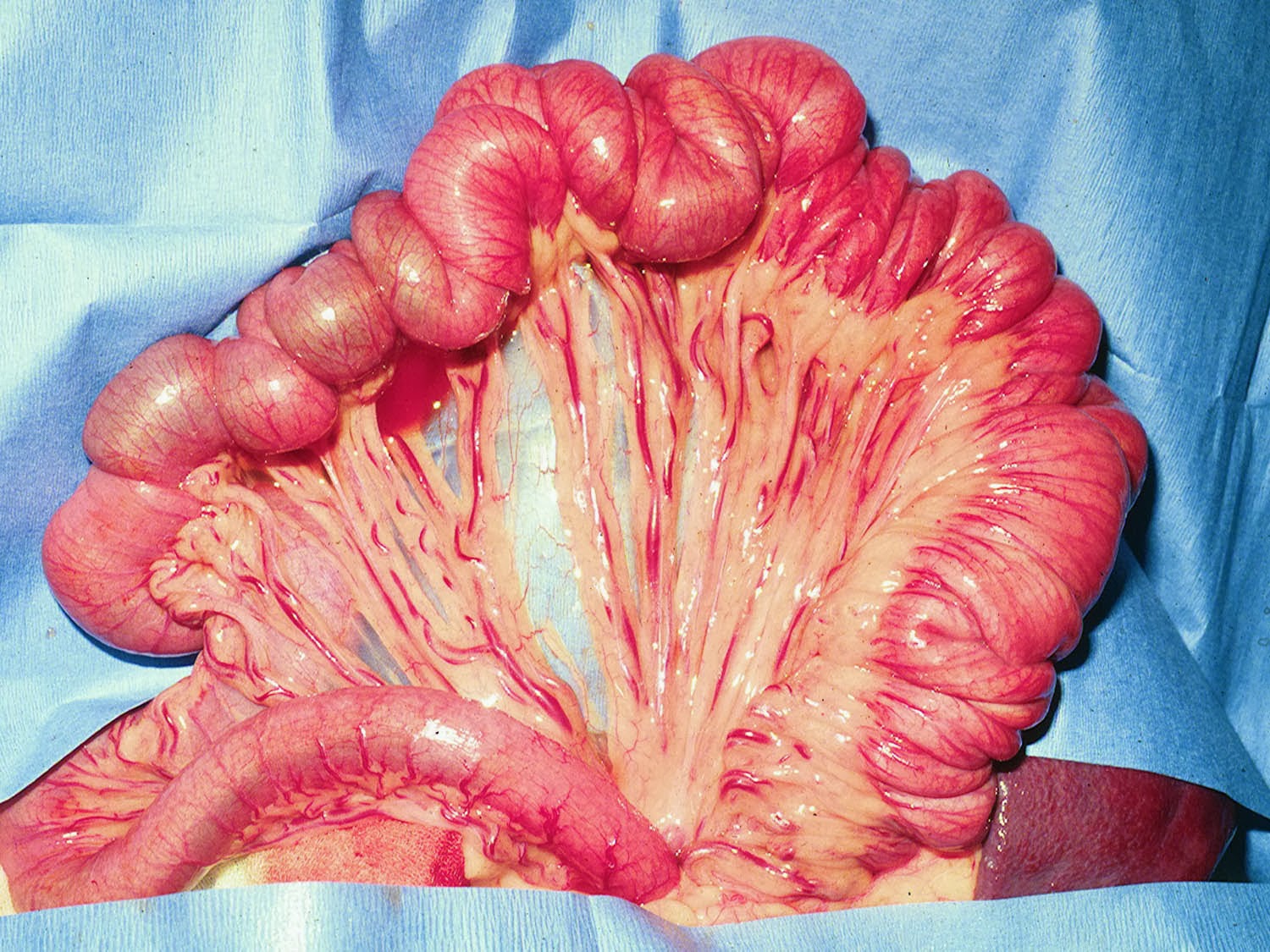 |
| Fig. 4: Intestinal plication in same dog as Fig. 2 radiographs |
 |
| Fig. 5: Intestinal plication in same dog as Fig. 3 |
If no foreign body is present in the stomach make the enterotomy in the middle of the plicated bowel. I find that many times the entire linear foreign body can be removed through one enterotomy if you patiently apply gentle traction on the foreign body while gradually releasing the plication of the intestine with your other hand or with the assistant’s help.(Fig. 6) Use suction and abdominal sponges to prevent peritoneal contamination.
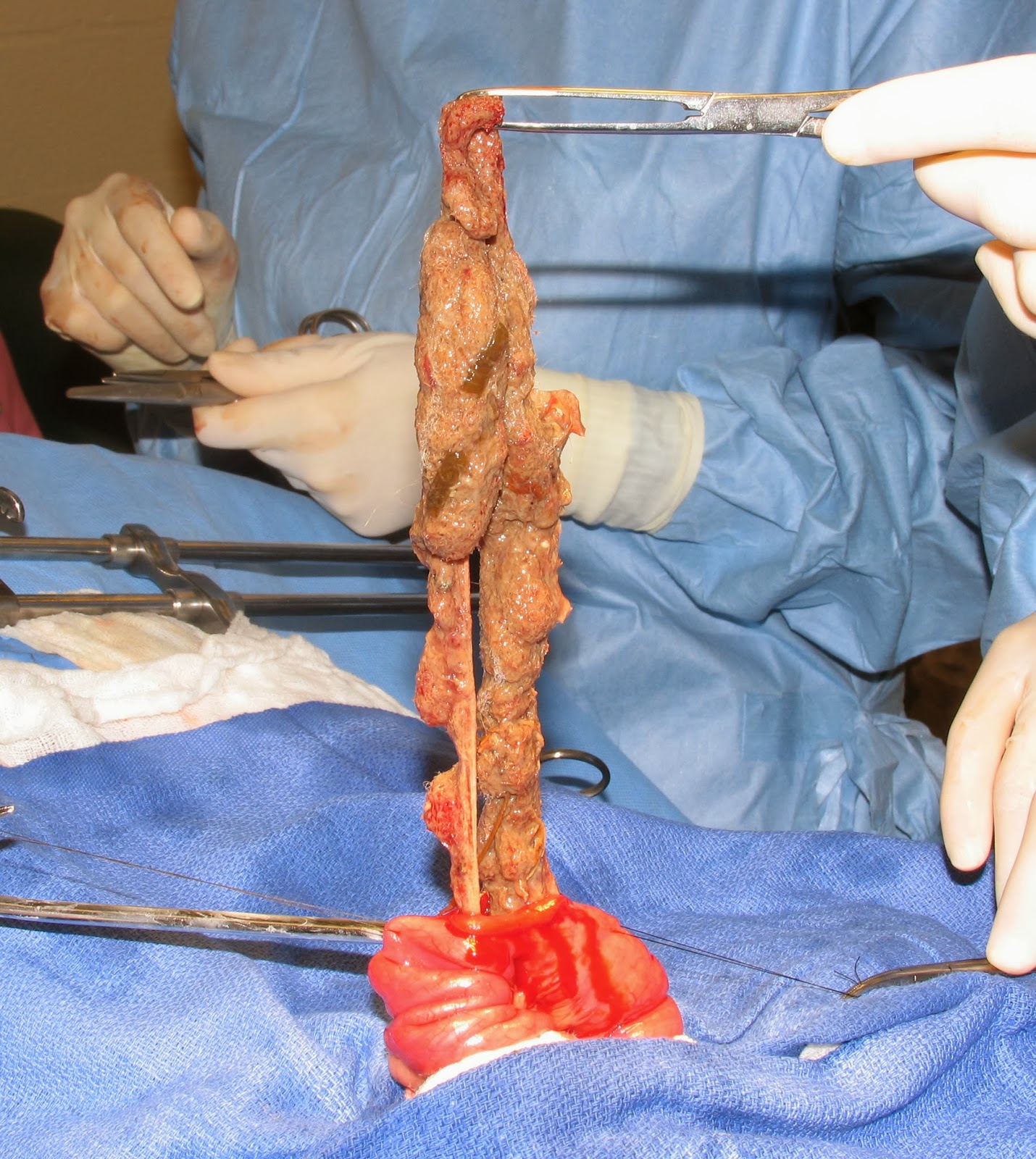 |
| Fig. 6: Linear foreign body removed through one enterotomy. |
String foreign bodies in cats are usually present in the stomach
and intestine. If so, begin with a gastrotomy to remove that portion of the
string. (See previous blog on gastrotomy) Gently pull the foreign body through
the gastrotomy in attempt to remove the intestinal portion as well. If that is
not possible remove as much as possible and then cut the string and close the
gastrotomy. Remove the remainder of the string through one or more enterotomy
incisions. (Fig. 7)
 |
| Fig. 7: String foreign body in a cat being removed through an enterotomy |
Also, prior to
performing the enterotomy attempt to milk the foreign material into one
segment of the intestine to make it easier to remove via a single enterotomy. Multiple
incisions in the gastrointestinal tract were one risk factor for higher
mortality in a large study of dogs and cats with intestinal foreign bodies.2
Perform resection and anastomosis of bowel that has been
perforated. Do not try to simply close the perforations; the tissue is not
healthy and normal healing is unlikely. Try to avoid multiple anastomoses of
the bowel; if possible include all the perforations in one resected segment so
that only 1 anastomosis results.
If bowel perforation is present obtain samples of peritoneal
fluid for culture, flush the abdomen with copious amounts of sterile saline,
and place a closed suction drain (e.g. Jackson-Pratt drain).
Postoperative Care
See blog on intestinal biopsy for routine care of intestinal
surgery patients. Postoperative care of animals with peritonitis will be
covered in a future post. Animals with linear foreign bodies have a guarded
prognosis compared to discrete, non-linear foreign bodies.
References
1.
Sharma A, Thompson MS, Scrivani PV, et.al. Comparison of radiography
and ultrasonography for diagnosing small-intestinal mechanical obstruction in
vomiting dogs. Vet Radiol Ultrasound.
2011 May-Jun;52(3):248-55
2.
Hayes G. Gastrointestinal
foreign bodies in dogs and cats: a retrospective study of 208 cases. J Small Anim Pract. 2009 Nov;50(11):576-83.
No comments:
Post a Comment