Author

Stephen J. Birchard DVM, MS, Diplomate ACVS
Showing posts sorted by relevance for query axial pattern flap. Sort by date Show all posts

Tuesday, April 1, 2014

Large Skin Defects in Dogs and Cats Treated With Axial Pattern Flaps (And one case where puppies are nursing from the leg!)

Fig. 1: Diagram of basic steps for caudal superficial epigastric axial pattern skin flap.
From: Birchard SJ, Smeak DD. Selected skin graft and reconstructive techniques. In: Saunders Manual of Small Animal Practice. 3rd edition, Birchard and Sherding, eds., Elsevier, St. Louis, 2006. Pg 557

Axial pattern flaps are skin flaps that are developed using a major direct cutaneous artery as the primary blood supply.  Considerably more flexibility in the length and mobility of these flaps is possible compared to random skin flaps (e.g., the one used in Hershey; see blog from 2/24/14).  These flaps can be made with long vascular pedicles to transfer skin to more remote areas of the body compared to random flaps.  This is because of preservation of the direct cutaneous artery allowing adequate perfusion of a large area of tissue.  
Axial pattern flaps can be based upon many direct cutaneous arteries, such as the caudal superficial epigastric, cervical cutaneous branch of the omocervical, thoracodorsal, deep circumflex iliac, and the genicular branch of the saphenous artery.  The caudal superficial axial pattern flap is commonly used for large defects of the proximal thigh and flank.  The thoracodorsal axial pattern flap is used for defects of the distal humeral area or elbow. These 2 techniques will be described here.  However, other axial pattern flaps as listed above can be created using similar principles and are described in current veterinary surgical textbooks.

Indications
Indications for axial pattern flaps are similar to random subdermal flaps except that these flaps are used when the skin defect is very large. Large skin defects that occur due to trauma, mass excision, or other etiologies resulting in a loss of skin could benefit from this reconstructive technique. A major direct cutaneous artery and vein must be fairly close to the skin defect for this technique to be indicated.

Surgical Procedures
Caudal Superficial Epigastric Axial Pattern Flap
Use similar preparation and tissue handling principles described for advancement or rotation flaps. Be particularly careful when positioning the patient such that the vascular pedicle will not become distorted before planning your incisions.  Incisions are created as shown in Figure 1.  The mammary chain up to and including the 3rd gland can be included.  Deeply undermine the flap just superficial to the abdominal fascia. Dissect very carefully around the origin of the direct cutaneous vessel to avoid inadvertent damage to this vessel that is vital to the survival of the flap.  Avoid creating a kink in the base of the flap that could obstruct blood flow. (Fig. 2)
Fig. 2: Caudal superficial epigastric axial pattern flap to close a medial  thigh wound
Drain the dead space if needed with a closed suction drain and suture the flap to the defect as described for the other flap techniques. Close the donor site after closing the flap.
Fig. 3: Healed caudal superficial flap on the stifle of a dog. Note nipples that have been transposed with the flap.
Fig. 4: Caudal superficial axial pattern flap was performed on this in tact female dog that subsequently got pregnant. Puppies are nursing from the transposed mammary glands on the leg. From: Pavletic MM. Axial pattern flaps in small animal practice. Vet Clin North Am Small Anim Pract. 1990 Jan;20(1):105-25.
Thoracodorsal Axial Pattern Flap
Fig. 5: Diagram of basic steps for thoracodorsal axial pattern flap.
From: Birchard SJ, Smeak DD. Selected skin graft and reconstructive techniques. In: Saunders Manual of Small Animal Practice. 3rd edition, Birchard and Sherding, eds., Elsevier, St. Louis, 2006. Pg 557
A rectangular flap of skin is created based on the thoracodorsal artery (Fig. 6) 
Fig. 6: Thoracodorsal flap planned for this non-healing wound secondary to tumor removal followed by radiation therapy. Four previous attempts to close thewound had been performed. 
Lines indicate proposed incisions; X marks the origin of the thoracodorsal artery.
The flap is undermined deep to the cutaneous trunci muscle and rotated to cover the defect. A bridging incision is made if necessary to allow suturing the flap to the skin proximal to the defect. The flap is rotated to the defect and sutured in place. (Fig. 7)
Fig. 7: Same dog as figure 5 with completed flap
Fig. 8: Same dog as figure 5, 2 weeks postoperatively
Care and Complications (All flap types)
Restrict exercise until suture removal. Apply an Elizabethan collar before the patient is recovered from anesthesia and leave on the animal until the flaps are completely healed. Place a well-padded bandage on the front leg after thoracodorsal flaps are used to cover the elbow. Pressure necrosis of the potion of flap directly over the elbow can occur if not protected and cushioned. Change wound dressings as necessary.
Major complications resulting from skin flaps include local wound problems such as:  partial or total ischemia of the flap, infection, seroma, and dehiscence of the flap or donor suture line. Mild bruising of the flap for the first few days postoperatively is expected. Severe discoloration with a sharp demarcation between normal and abnormal skin is likely indicating flap ischemia and impending necrosis. Dehiscence of donor site incisions is usually due to excessive skin tension.  If dehiscence occurs, allow these areas to heal by second intention.

References

  1. Birchard SJ, Smeak DD. Selected skin graft and reconstructive techniques. In: Saunders Manual of Small Animal Practice. third edition, Birchard and Sherding, eds., Elsevier, St. Louis, 2006. Pg 557
  2.  Aper R1, Smeak DD. Complications and outcome after thoracodorsal axial pattern flap reconstruction of forelimb skin defects in 10 dogs, 1989-2001. Vet Surg. 2003 Jul-Aug;32(4):378-84.
  3. Aper RL1, Smeak DD.Clinical evaluation of caudal superficial epigastric axial pattern flap reconstruction of skin defects in 10 dogs(1989-2001). J Am Anim Hosp Assoc. 2005 May-Jun;41(3):185-92.
  4. Pavletic MM. Axial pattern flaps in small animal practice. Vet Clin North Am Small Anim Pract. 1990 Jan;20(1):105-25.


Saturday, April 21, 2018

Axial Pattern Flap Reconstruction of a Chronic Non-Healing Elbow Wound in a Golden Retriever

Ginger is a 7-year-old female spayed golden retriever who presented to MedVet Toledo with a chronic non-healing open skin wound over the right elbow. (Fig. 1) 

Fig. 1: Ginger
The owner adopted Ginger 1.5 years prior to presentation and the wound was present at that time. Ginger was otherwise healthy with no major medical issues. Serous fluid drained from the wound occasionally. Antibiotic therapy previously prescribed did not result in significant improvement of the wound.

Physical examination of Ginger was normal except for a large (8cm diameter) open wound over the right elbow that appeared to be a decubital ulcer. (Fig. 2) 
Fig. 2: The non-healing chronic open wound over the right elbow on Ginger
Extensive fibrotic scar tissue was present around the periphery of the circular wound, and unhealthy granulation tissue was seen in the wound center. Little to no discharge was present on the wound surface.

A fine needle aspirate of the peripheral scar tissue was performed to rule out neoplasia and the cytology results indicated chronic inflammation. Routine preoperative CBC and serum chemistry profile were normal. The plan was to anesthetize Ginger, perform an extensive surgical debridement of the fibrotic peripheral tissues, and reconstruct the skin using an axial pattern skin flap. 

Ginger was anesthetized and the right elbow and surrounding skin clipped and prepared for aseptic surgery. The donut shaped abnormal scar tissue was removed using blunt and sharp dissection. (Fig. 3) 
Fig. 3: The right elbow of Ginger immediately after surgical debridement of the chronic wound. 
During this dissection it became clear that the tissue was highly vascularized. Hemostasis was difficult and bleeding became brisk throughout the dissected area. The hemorrhage was controlled with electrocautery and direct pressure. We elected to delay the skin flap reconstruction so that open wound management with tie-over bandages could be used on the elbow to allow the debrided area to be in more optimal condition for the definitive reconstruction procedure. (for more information see: tie-over bandages)

One week after the surgical debridement Ginger was re-admitted to the hospital for the skin flap procedure. Ginger was placed under general anesthesia and a thoracodorsal axial pattern skin flap was performed to reconstruct the elbow decubitalulcer. (Fig. 4)
(for more information see: axial pattern skin flaps) 
Fig. 4: Completed thoracodorsal axial pattern flap reconstruction of the elbow on Ginger.
White arrow indicates direction of transfer of the rectangular skin flap harvested from skin over the scapula.
Complete coverage of the wound was achieved with the skin flap. A spica bandage was placed over the leg and thorax to protect the incisions and Ginger was recovered from anesthesia. 

The following day Ginger’s bandage was changed and she was discharged from the hospital. Bandage changes occurred every 2-3 days until the incisions were healed and staples removed. (Fig. 5) 
Fig. 5: Appearance of the surgical incisions on Ginger 10 days postoperatively.
Ginger finally was discharged with no rechecks necessary unless problems arose. However 5 months later she represented to MedVet Toledo for problems unrelated to her elbow. The owner reported that since the skin flap reconstruction Ginger had been much more comfortable and mobile. Ginger’s skin flap had grown a thick, bushy amount of hair. There was redundant skin at the elbow but the overall cosmetic appearance was quite satisfactory. (Fig. 6) In general the owner was very pleased with the end result of the reconstruction.
Fig. 6: Appearance of Ginger's right elbow area 5 months after reconstruction.
Chronic wounds of the elbow in dogs are one of the most difficult to manage. Skin tension, excessive motion, and trauma to the area combine to interfere with healing in this part of the body. Debridement and local advancement of skin over the defect is generally unrewarding for reconstruction, particularly if it results in a suture line directly over the olecranon. Skin flap techniques that bring healthy, robust tissue into the site without creating a suture line over the olecranon, such as an axial pattern flap, is more likely to be successful.

Several aspects of the postoperative care are key to the success of this procedure. A well-padded bandage to protect and cushion the skin flap is mandatory for at least 10-14 days postoperatively. Exercise restriction is also important along with frequent postoperative rechecks to monitor for flap survival and healing. 

One clinical study of 10 dogs having thoracodorsal axial pattern flaps found complications to be common but good functional and cosmetic outcome resulted in 6 of 7 dogs long term follow up. (1)

References

1. Aper R, Smeak, D:  Complications and Outcome After Thoracodorsal Axial Pattern Flap Reconstruction of Forelimb Skin Defects in 10 Dogs, 1989–2001 32:4, 2003, Pages 378-384.

Tuesday, June 28, 2022

Medical and Surgical Treatment of Severe Thermal Burns in a Cat


Signalment and Presentation

Maximus, a healthy 6-month-old mixed breed neutered male cat, was presented for severe thermal burns over large areas of his body after jumping into a bathtub filled with scalding hot water.  The injury occurred just a few hours before presentation.


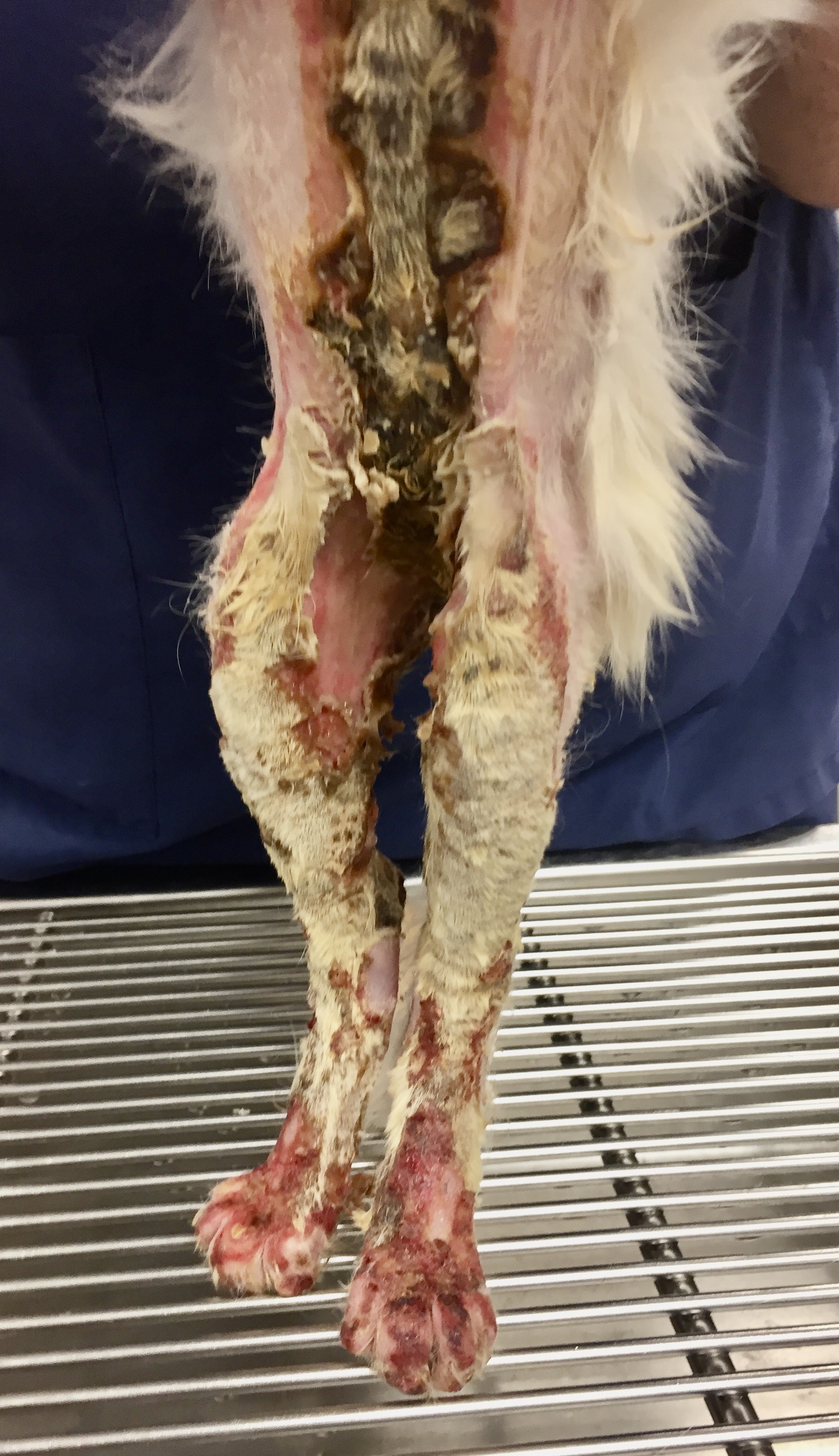
On admission, Maximus was tachycardic and severely painful. All four paws were red, swollen, and bleeding. The skin on his ventral abdomen and thorax was bright red and bruised. It appeared that the injury affected about 50% of his body. There was no other previous history of medical problems.


Initial Treatment

Maximus was initially administered intravenous fluids, antibiotics, and buprenorphine for pain. Continuous intravenous infusion of fentanyl was added to the treatment regimen soon after admission. Blood tests showed evidence of dehydration and elevated serum potassium. Silver sulfadiazine was used as the topical cream medication on the burned areas.


Maximus was non-ambulatory because the skin of his footpads was peeling off, leaving subcutaneous tissue exposed. Over the next several hours, the burned areas on his legs, chest, and belly gradually became purple in color. Some of the burned skin on his chest was becoming hard and leathery. (Fig. 1a,b) On a pain scale from 1-24 (1 being little or no pain, 24 being the worst pain possible), the attending clinician listed Maximus as 24. Intravenous lidocaine and ketamine were added to the fentanyl infusion to provide more analgesia. The dosage of fentanyl was also increased.

Fig. 1a: Maximus several hours after admission to the hospital

Fig. 1b


After sedating him with intravenous propofol, all of Maximus’ burned areas were carefully clipped and cleaned. With the hair removed, the burned regions were more clearly visible; the total surface area of his burned body appeared to be closer to 60%. 


More blood tests showed that Maximus’ albumin was already below normal. Albumin was being depleted because of the severe inflammation of the damaged skin and loss of albumin-rich fluid that oozed from his wounds. A nasogastric tube was placed to augment his dietary intake. 


Wound Management

On day two after admission, Manuka honey (Medihoney) was begun as the topical medication on the wounds, and standard bandaging was continued. The burned areas were gradually declaring themselves, and on day 4, some were starting to turn yellowish-brown and becoming hard and crusty. Escars were developing, indicating full-thickness skin injury. (Fig. 2)

Fig. 2: Escar development over mid abdomen and rear legs


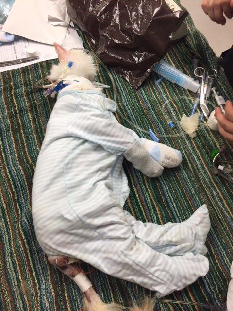
To simplify the bandage changes that were time-consuming and painful, the attending clinician Dr. Becky Ball began covering Maximus with infant onesies, little pajama outfits that completely covered the entire body, legs, and feet. (Fig. 3) Each day the onesie was removed, the wounds gently cleaned, Medihoney ointment applied, and a clean onesie replaced. 

Fig. 3: Onesie pajama bandage applied.


Maximus was discharged 12 days after admission. The owner, a physician, assumed the nursing care at home. Rechecks with Dr. Ball were scheduled every 4-5 days for wound assessment. The escars gradually peeled away from the healthy skin, exposing new granulation tissue underneath. No evidence of infection was seen, and Maximus' appetite was good. 


Maximus continued to be re-examined about every two weeks. The eschars completely sloughed off, and the skin defects were contracting and closing well. But, as the skin tightened in his inguinal region, scar tissue pulled his rear legs medially, severely restricting his mobility. (Fig. 4) Surgery would be necessary to alleviate the contracture and allow Maximus to use the hind legs better. 

Fig. 4: Wound contraction of inguinal skin.

Skin Reconstruction


Seven months after Maximus’ injury, a caudal superficial epigastric axial pattern skin flap was performed to alleviate the inguinal wound contracture. (Fig. 5a-b) Click here for a detailed description of the skin flap procedure. The surgery went well, and he was discharged from the hospital the following day.

Fig. 5a: Axial pattern skin flap design. Arrow indicates the direction of rotation of the flap after it is dissected out.

Fig. 5b: Completed skin flap procedure.


Seven days postoperatively, suture removal was performed. The skin flap was viable and had healed well to adjacent skin. Maximus was much more mobile and less painful. Seven months postoperatively, he was examined again and was doing well with all wounds healed and his function back to normal. (Fig. 6, 7)

Fig. 6: Appearance of inguinal area seven months after skin flap reconstruction.

Fig. 7: Maximus and his owner 15 months after his injury.


Discussion


Maximus overcame a severe injury with burns over a large portion of his body. He exemplifies the remarkable wound healing process, particularly when supported with good nursing care. Manuka honey was effective in preventing infection and encouraging wound healing. Dietary support was instrumental in avoiding hypoproteinemia and malnutrition. Excessive wound contracture necessitated skin flap reconstruction, improving mobility and relieving chronic pain. 


Maximus' owner, a neonatologist, was instrumental in his recovery. She assumed the challenging nursing care at home and was diligent in the tedious work of medicating, bandaging, and maintaining good hygiene.


Maximus’ complete story is told in Dr. Birchard’s book: Their Tails Kept Wagging: Pets Show Us How Hope, Forgiveness, and Love Prevail, a collection of inspirational stories of severely ill pets who survived due to modern medicine and compassionate caregivers. For more information, click here.


Tuesday, November 24, 2015

Complete Surgical Excision of Mast Cell Tumor in Dogs and Cats

A wide variety of skin tumors occur in dogs and cats, both benign and malignant.  An important principle of surgical management of these tumors is to establish a diagnosis before the operation.  Fine needle aspiration (FNA) is a practical and reasonably accurate method to biopsy skin masses, and the results allow clinicians to plan appropriate treatments.  For example, benign skin tumors such as epidermal inclusion cysts require only a marginal excision and routine skin closure.  Malignant skin tumors, such as mast cell tumor (MCT), require extensive tissue resection (e.g. removal of 2-3 cm of normal tissue with the mass) followed by more complicated reconstruction in some cases.  An adequate deep margin of normal tissue should be removed with the tumor as well as medial and lateral margins. Excise a section of the tissue layer below the tumor to achieve a complete resection. If the tumor is located in the subcutaneous space remove the muscle or deep fascia below the tumor.

Preoperative Considerations
As is true for any animal with neoplasia, tumor staging is done prior to surgery to establish the extent of disease. Appropriate imaging, such as thoracic radiographs and abdominal ultrasound, is used to examine for metastasis or other unrelated problems. Also carefully examine regional lymph nodes and if enlarged perform FNA. With MCT, administer preoperative antihistamines such as diphenhdyramine to reduce the inflammation associated with histamine release by the tumor. The drug can be given either orally for 1 or more days preoperatively, or parenterally just before surgery. Avoid excessive manipulation of the tumor before surgery to prevent degranulation of mast cells and release of histamine.

Be sure to warn owners about potential complications of surgical removal of MCT. Even with antihistamine pre-treatment wound complications such as excessive inflammation, seroma, and dehiscence are possible.

Surgical Technique
After placing the dog or cat under general anesthesia, perform a wide aseptic preparation of the surgical site. (Fig 1) 
Fig. 1: Cutaneous mast cell tumor (circle) over the dorsal thoracic area in a spaniel.
(note Figs 3-7 are the same dog as in this picture)
Use a sterile ruler and marking pen to delineate the mass (Fig 2), then draw a circle around the tumor that is 2-3 cm from the edge of the mass.(Fig 3)
Fig. 2: Sterile surgical marking pen and ruler to map surgical margins around skin tumors.
Fig. 3: MCT (inner circle and X) delineated by an outer circle of 2cm margins of normal skin
Draw lines that taper the incision on each end to make the incision an ellipse and thus avoid having “dog ears” of skin on the ends.(Fig. 3) Be sure that the long axis of the resultant incision is parallel to the tension lines in that area of the body.

Make the incisions on the proposed lines and continue the dissection into the deep tissues. Avoid dissecting toward the tumor; as you proceed deeper in the tissues continue to honor the 2 or 3 cm margin originally mapped on the skin. Once the desired layer of deep margin has been reached, incise the fascia or muscle, lift the tumor and associated tissue (en bloc), and dissect the block of tissue completely free.(Fig. 4)
Fig. 4: Intraoperative picture with skin mass and underlying tissue
being removed from right to left. Note underlying muscle being removed with the mass.
Wide excision of skin or subcutaneous masses frequently leaves large skin defects that can be difficult to close. When primary closure cannot be obtained due to excessive skin tension, consider either immediate or staged flap or graft reconstruction. (see blogs from 3/20/14 on punch skin grafts and 4/1/14 on axial pattern skin flaps) If local tissues are adequate for closure, use the “Rule of Halves” technique (see blog from 11/3/14 on closure of elliptical skin incisions). A towel clamp can be used to initially bring the skin together at the middle of the incision to make suture placement easier.(Fig. 5) 
Fig. 5: A towel clamp is used to temporarily relieve tension and allow
suture closure the incision.
Close deep tissues at the middle of the incision first, then continue to place sutures in the rule of halves manner to achieve complete closure.(Fig. 6, 7)  
Fig. 6: Deep sutures have been placed in the middle of the incision; the next 2 deep sutures
will be placed at the arrows in the "Rule of Halves" manner.

Fig. 7: Final appearance of closed incision
Place a closed suction drain if excessive dead space exists in the deep tissue layers that cannot be closed (see blog from 3/15/14 on Jackson Pratt drains)

After removal of the mass, ink the tissue specimen with appropriate dye such as India ink to allow the pathologist to identify the margins of the excision. Also, place a suture on either the cranial or caudal aspect of the specimen to further orient the pathologist in case one of the margins shows incomplete excision of the tumor.

Postoperative Care
Routine supportive care is administered after removal of mast cell tumors. Avoid NSAIDS administration on MCT dogs to prevent compounding the gastric irritation from the histamine. Tramadol is a good alternative postoperative analgesic. Monitor the surgical incision for swelling or bleeding, and instruct owners to limit exercise and monitor the surgical site carefully at home. Although most dogs and cats recover without major systemic complications after removal of MCT, systemic vascular collapse is possible from massive histamine release in rare cases. Fluid resuscitation and corticosteroid administration may be necessary to support and stabilize the patient if this occurs.

Prognosis
Long-term outcome is dependent upon the histopathologic classification of the MCT. There are 2 major classification schemes now used by pathologists, i.e., Grade 1, 2 and 3 (1 is low grade, 3 is high grade and 2 is intermediate grade) or a simpler 2 tier system of low-grade vs. high grade.(1) Regardless of the system used, the higher the grade of MCT the poorer the prognosis.(1,2) Information on adjunctive therapy such as chemotherapy or radiation therapy is readily available and may be useful in animals with incompletely excised or metastatic tumors.(3)

References

1. Sabattini S, Scarpa F, Berlato D, Bettini G. Histologic grading of canine mast cell tumor: is 2 better than 3? Vet Pathol. 2015 Jan;52(1):70-3.
2. Donnelly L, Mullin C, Balko J, et.al. Evaluation of histological grade and histologically tumour-free margins as predictors of local recurrence in completely excised canine mast cell tumours.Vet Comp Oncol. 2015 Mar;13(1):70-6.
3. London CA, Thamm DH. Mast cell tumor. In: Small Animal Clinical Oncology, eds: Withrow S, MacEwen G, Elsevier, 2013, pg. 335.